itnTV

视频:The Rise of Point of Care Ultrasound During COVID-19

Arun Nagdev, M.D., director of emergency ultrasound at theAlameda Health System, clinical associate professor, University of California San Francisco (UCSF) School of Medicine, and incoming president for theAmerican College of Emergency Physicians(ACEP) ultrasound section, explains the rise inpoint-of-care ultrasound(POCUS) since the start of theCOVID-19pandemic. Nagdev has used cart-based and several hand-held systems to the emergency department to triage patients, help sort out COVID and non-COVID patients and identify patients that need more detailed radiology imaging or with issues that need immediate attention, such as pulmonary embolism, heart attacks and aortic aneurisms.

Nagdev also serves as senior director of clinical education for start-up POCUS companyExo该公司正在开发一种新的基于芯片的手持超声系统,配有高清晰度换能器。

Watch the related视频:How to Image COVID-19 and Radiological Presentations of the Virus— Interview with Margarita Revzin, M.D.

More COVID-19 content can be found here

Radiology Imaging

Point-of-Care Ultrasound (POCUS)| May 13, 2022

Arun Nagdev, M.D., director of emergency ultrasound at theAlameda Health System, clinical associate professor, University of California San Francisco (UCSF) School of Medicine, and incoming president for theAmerican College of Emergency Physicians(ACEP) ultrasound section, explains the rise inpoint-of-care ultrasound(POCUS) since the start of theCOVID-19pandemic. Nagdev has used cart-based and several hand-held systems to the emergency department to triage patients, help sort out COVID and non-COVID patients and identify patients that need more detailed radiology imaging or with issues that need immediate attention, such as pulmonary embolism, heart attacks and aortic aneurisms.

Nagdev also serves as senior director of clinical education for start-up POCUS companyExo该公司正在开发一种新的基于芯片的手持超声系统,配有高清晰度换能器。

Watch the related视频:How to Image COVID-19 and Radiological Presentations of the Virus— Interview with Margarita Revzin, M.D.

More COVID-19 content can be found here

Computed Tomography (CT)| March 09, 2022

AtRSNA 2021, Philips highlighted the launch of two new innovativeCT systems– the multi-energySpectral CT 7500and theCT 5100 Incisivewith embeddedAIcapabilities.ITNspoke with Wendy Winkle Lawless, CT Business Market Leader - North America, Philips, to learn more about these new systems.

Digital Radiography (DR)| March 01, 2022

AtRSNA 2021, Konica Minolta introduced themKDR XpressMobile X-ray system and theAero DR Carbon flat panel detector.Also on display wasnVoq’s cloud-based speech recognition and automation solution and new features for theExa platformthat automate common clinical and administrative tasks.

You can find more RSNA21 content here

Contrast Media| February 23, 2022

With the use ofcontrast agentsandradiotracerson the rise,GE Healthcarehas seen increases in demand across their pharmaceutical diagnostics business. AtRSNA 2021, Marco Campione, General Manager, Pharmaceutical Diagnostics Americas, GE Healthcare, shared how recent investments are helping to meet this increased demand.

Find more RSNA21 content here

Magnetic Resonance Imaging (MRI)| February 09, 2022

AtRSNA 2021, Fujifilm launched its newVelocity MRIsystem. It is designed to streamline workflow and enhance the patient experience with its open gantry, integrated RF coils and reconstruction techniques. Shawn Etheridge, Director, CT and MRI Marketing forFujifilm, unveiled the system on the RSNA21 show floor.

Fujifilm Launches the Velocity MRI System at RSNA 2021

Additional RSNA21 content

Quality Assurance (QA)| January 31, 2022

Sun Nuclearhighlighted two resources forCT Quality AssuranceatRSNA 2021ITNvisited their booth to learn more about theMercury 4.0 Phantomand theMulti-Energy CT Phantomfrom Thomas Webb, Global Product Marketing Manager

Find more RSNA news and videos

Digital Radiography (DR)| January 31, 2022

Agfa recently announced the launch of the newValorydigital radiography room, which was designed for health care facilities requiring a highly productive radiography solution.

ITNstopped by theAgfabooth atRSNA 2021to learn more about this new system fromGeorges Espada, Head of Digital Radiography, and Karol Wesolowski, Global Category Leader.

Coronavirus (COVID-19)| January 31, 2022

Marc Succi, M.D., an emergency radiologist at MGH and executive director of the MESH Incubator, an in-house innovation and entrepreneurship center, and Ottavia Zattra, a fourth-year medical student at Harvard Medical School, explain a study they authored showing there might be higher cancer rates due to lower numbers of CT scans duringCOVID-19.They presented this study as a late-breaker at theRadiological Society of North America (RSNA)2021 annual meeting.

COVID caused many people to delay seeing their doctors. Their study found a corresponding 82% drop in CT imaging for cancer screening. CT is also used for initial cancer workups, to monitor active cancer and post procedure surveillance, which all also showed decline since the start of the pandemic.

Read more about the studyCOVID-19 Fallout May Lead to More Cancer Deaths

Computed Tomography (CT)| January 28, 2022

Charlie Hamm, M.D., a radiology resident at the Charité University Hospital of Berlin, Germany, presented a late-breaking study at theRadiological Society of North America (RSNA)2021 annual meeting on the use ofcomputed tomography (CT)scans to investigate dinosaur bones non-destructively. In the process of examining a tyrannosaurus rex jaw bone that is more than 66 million years old, a bone tumor was found and clearly shown on the CT scans.

This feasibility study to determine if CT can be used to aid paleontology was done in collaboration with the Museum für Naturkunde Berlin. Dual-energy computed tomography (DECT) was used to provide information about tissue composition and disease processes not possible with single-energy CT.

Read more in the articleCT Uncovers Bone Disease in Tyrannosaurus Rex Jaw

Find more RSNA news and video

Quality Assurance (QA)| January 28, 2022

Mahadevappa Mahesh, Ph.D., chief physicist, Johns Hopkins Hospital, and professor of radiology and radiological science, explains the basics involved inquality assurance (QA)of radiology imaging systems. He spoke toITNat theRadiological Society of North America (RSNA)2021 annual meeting.

He explains the role of the medical physicist in keeping X-ray imaging systems such as CT, angiography and mammography calibrated and checking the device output of radiation. This is performed by imaging phantoms that mimic a simplified representation of the human body.

Artificial intelligence (AI)use is growing in imaging and medical physics and QA of these systems might also become a duty of the medical physicist in some AI imaging applications.

Find more content on QA systems

Find more RSNA news and video

Computed Tomography (CT)| January 27, 2022

Cynthia McCollough, Ph.D.,director of MayoClinic's CT Clinical Innovation Center,explains how photon-countingcomputed tomography (CT)detectors work and why it is a better technology over conventional CT systems. She helped Siemens develop the Naeotom Alpha, the first photo-counting CT system to be approved by the FDA in the fall of 2021. She spoke toITNat theRadiological Society of North America (RSNA)2021 annual meeting.

Read more about the first commercial photon-counting scanner

The device uses the emerging CT technology of photon-counting detectors, which can measure each individual X-ray photon that passes through a patient's body, as opposed to current systems which use detectors that measure the total energy contained in many X-rays at once. By "counting" each individual X-ray photon, more detailed information about the patient can be obtained and used to create images with less information that is not useful, such as image noise.

目前的CT技术采用两步转换过程,利用探测器中的闪烁层将x射线光子转换为可见光。然后,光电二极管光传感器将可见光转换成数字信号。由于这一中间步骤,关于x射线能量的重要信息丢失了,不能再用于诊断。此外,对比度降低,图像不那么清晰。

Photon-counting detectors use a single step of direct conversion of X-rays into electrical current, and skips the step of converting X-rays into visible light. This allows the energy thresholds of each pulse to be collected and binned based on different kilovolt (kV) energy levels. This creates data to improve contrast and enable dual-energy, spectral imaging. The direct conversion also helps improve image quality without information loss. This improves image sharpness and contrast.

Photon-counting detectors have already been used for several years in high-energy physics and nuclear imaging. However, these previously generation photon-counting detectors could not be used with a clinical CT scanner because they could not keep up with the high higher rate of photons reaching the detector. The detector on the Naeotom Alpha was designed for this increased speed.

Related Photon-counting CT Content:

Mayo Clinic Begins Use of Third-Generation Photon-counting CT Clinical Research Detector

视频:New Advances in CT Imaging Technology— Interview with Cynthia McCollough, Ph.D.

视频:Photon Counting Detectors Will be the Next Major Advance in Computed Tomography— Interview with Todd Villines, M.D.

Key Trends in Cardiac CT at SCCT 2020

GE Healthcare Pioneers Photon Counting CT with Prismatic Sensors Acquisition

Top Trend Takeaways in Radiology From RSNA 2020

NeuroLogica Joins Forces with Massachusetts General Hospital to Pilot Photon Counting CT at the Patient’s Point-Of-Care Using OmniTom Elite CT

视频:Advances in Cardiac CT Imaging— Interview with David Bluemke, M.D.

Computed Tomography (CT)| January 27, 2022

Cynthia McCollough, Ph.D.,director of MayoClinic's Computed Tomography (CT) Clinical Innovation Center,explains how CT dose tracking software works and offers advice to centers that record this patient level and device information. She spoke to ITN at theRadiological Society of North America (RSNA)2021 annual meeting.

Dose tracking software allows hospitals and imaging centers to track what levels of radiation they are using by exam type protocol. It can show technologists who are using higher than required doses that may need additional ALARA training. The radiation dose tracking systems also can help track the amount of radiation a patient has received over time.

Related Radiation Dose Tracking Systems:

Disputed EHR Dose Levels Could Keep Patients From Necessary Imaging Exams

Medical Imaging Radiation Exposure in U.S. Dropped Over Past Decade

视频:Radiation From Medical Imaging in U.S. Dropped Over Past Decade

The Basics of Radiation Dose Monitoring in Medical Imaging

视频:Radiation Dose Monitoring in Medical Imaging— Interview with Mahadevappa Mahesh, Ph.D.

----

Magnetic Resonance Imaging (MRI)| January 24, 2022

With the recent launch of theMagnifico Open, Italian companyEsaotehas entered the openMRIwhole body space.ITNhad a conversation withFranco Fontana, CEO of Esaote, atRSNA21

Magnifico Open, which adds to the range of Esaote products unveiled in 2021, is anopen magnetic resonance systemwith the latest technology. The wide choice of receiver coils and state-of-the-art MRI technology offer the user excellent image quality, while the permanent magnet makes it easy to use and lowers operating costs. The open magnet and the easy-to-access patient table also facilitate, speed up and make patient positioning more comfortable, ideal for both the claustrophobic and for children.

View more RSNA21 content here

Esaote North America Receives FDA Approval of the Magnifico Open MRI System

Magnetic Resonance Imaging (MRI)| January 18, 2022

Orlando Simonetti, Ph.D.,professor, cardiovascular medicine, worked with Siemens to help develop a new, lower-fieldmagnetic resonance imaging (MRI)system, the Magnetom Free.Max. It can scan patients that previously may have been contraindicated because of implantable medical devices. One of the first systems installed in the U.S. is atThe Ohio State University Wexner Medical Center.It has a much lower magnetic field and a larger patient opening, removing barriers to MRI imaging for many patients.

Simonetti and his colleagues developed new techniques to boost the signal-to-noise ratio in MRI machines, which allowed the creation of a machine with a lower magnetic field strength that still enables high quality images.

The system gained FDA clearance in July 2021and was featured by Siemens at the Radiological Society of North America (RSNA) 2021 meeting.

The interview and footage was provided by The Ohio University State University Wexner Medical Center.

Read more in the articlesNew FDA-approved MRI Expands Access to Life-saving ImagingandOhio State Researchers Help Design New MRI, Expanding Access to Life-saving Imaging

Related MRI Content:

Siemens Healthineers Announces First U.S. Installation of Magnetom Free.Max 80 cm MR Scanner

FDA Clears Siemens Healthineers Magnetom Free.Max 80 cm MR Scanner

Ohio State Researchers Help Design New MRI, Expanding Access to Life-saving Imaging

Enterprise Imaging| January 13, 2022

Steve Holloway, company director atSignify Research, explains the trends he has seen over the past couple years inenterprise imaging.他在2021年北美放射学会会议上对ITN发表了讲话。

Holloway shared how medical imaging systems are expanding to include all departments in healthcare system enterprises that generate data, images and waveforms, so these items can be stored in a central location, rather than disparate silos or in separate systems requiring multiple logins or specific workstations. Most of these systems are are web enabled or web based, allowing users to work from anywhere as long as they have an internet connection. Most enterprise imaging systems also use a web-based vendor neutral archive, allowing DICOM and non-DICOM images to be stored there. All of these features allow easier and faster access to patient information and images.

He said these systems are becoming more inclusive of ologies outside of radiology and cardiology. Most notably isdigital pathology, which was featured by many enterprise imaging vendors at RSNA 2021.

Enterprise imaging systems are also acceptingpoint-of-care ultrasound (POCUS), which has exploded in use over the past two years with COVID, Holloway said.

Find more RSNA news and video

视频:Trends in Radiology IT seen at RSNA 2021— Interview with Jef Williams, Paragon Consultants

视频:Artificial Intelligence Trends in Medical Imaging— Interview with Sanjay Parekh, Ph.D, from Signify Research

视频:Examples of Improved PACS Workflow to Aid Speed and Efficiency

视频:The New Normal of Home Workstations, Teleradiology and Remote Reading— Interview with Elizabeth Hawk, M.D.

Technology Report: Artificial Intelligence in Radiology 2021

Technology Report: Enterprise Imaging 2019

Coronavirus (COVID-19)| December 14, 2021

Jean Jeudy, M.D., professor of radiology and vice chair of informatics at the University of Maryland School of Medicine, presented a late-breaking study at the 2021Radiological Society of North America (RSNA)meeting on COVID-19 linked myocarditis in college athletes.

A small but significant percentage of college athletes withCOVID-19develop myocarditis, a potentially dangerous inflammation of the heart muscle, that can only be seen oncardiac MRI, according to the study Jeudy presented. Myocarditis, which typically occurs as a result of a bacterial or viral infection, can affect the heart’s rhythm and ability to pump and often leaves behind lasting damage in the form of scarring to the heart muscle. It has been linked to as many as 20% of sudden deaths in young athletes. The COVID-19 pandemic raised concerns over an increased incidence of the condition in student-athletes.

For the new study, clinicians at schools in the highly competitiveBig Ten athletic conferencecollaborated to collect data on the frequency of myocarditis in student-athletes recovering from COVID-19 infection. Conference officials had required all athletes who had COVID-19 to get a series of cardiac tests before returning to play, providing a unique opportunity for researchers to collect data on the athletes’ cardiac status.

37名运动员(2.3%)被诊断为COVID-19心肌炎,与普通人群心肌炎发病率持平。然而,没有临床症状的运动员患心肌炎的比例高得惊人。20例(54%)的COVID-19心肌炎患者没有心脏症状,也没有心脏检测异常。只有心脏核磁共振成像发现了这个问题。

Read more details in the articleCOVID-19 Linked to Heart Inflammation in College Athletes

Find more RSNA news and video

Related COVID-19 Imaging and Myocarditis Content:

Overview of Myocarditis Cases Caused by the COVID-19 Vaccine

COVID-19 Linked to Heart Inflammation in College Athletes— RSNA 2021 late-breaker

Cardiac MRI of Myocarditis After COVID-19 Vaccination in Adolescents

Large International Study Reveals Spectrum of COVID-19 Brain Complications - RSNA 2021 late-breaker

COVID-19 During Pregnancy Doesn’t Harm Baby’s Brain

视频:Large Radiology Study Reveals Spectrum of COVID-19 Brain Complications — Interview with Scott Faro, M.D.

FDA Adds Myocarditis Warning to COVID mRNA Vaccine Clinician Fact Sheets

Small Number of Patients Have Myocarditis-like Illness After COVID-19 Vaccination

2022世界杯篮球预选赛赛程| December 13, 2021

Stamatia Destounis, M.D., FACR, chief of the American College of Radiology (ACR) Breast Commission, managing partner, Elizabeth Wende Breast Care, Rochester, N.Y., explains some of the key trends in2022世界杯篮球预选赛赛程at the 2021Radiological Society of North America (RSNA)meeting.

She discusses the trends of 3D mammography seeing rapid growth, adoption of synthetic 2D breast images from the tomosynthesis datasets, contrast-enhanced mammography, and breast MRI to help women with dense breast tissue. Destounis also discusses the use of artificial intelligence (AI) to help radiologists with finding what they needs with larger datasets in 3D mammography, and to help act as a second set of eyes.

Early in 2021, with the roll out of the COVID vaccines, one of the biggest headlines in radiology was that the vaccine can show false positives for cancer because it may cause inflammation of lymph nodes. Destounis explains this issue and how women's health centers have largely overcome this by asking patients about their vaccination status and planning imaging around the vaccination dates.

Related Breast Imaging Content:

COVID-19 Vaccine Can Cause False Positive Cancer Diagnosis

Help Spread Awareness of Potential COVID-19 Vaccine Imaging Side-effects

视频:COVID Vaccine May Cause Enlarged Lymph Nodes on Mammograms— Interview with Constance "Connie" Lehman, M.D.

COVID-19 Vaccination Axillary Adenopathy Detected During Breast Imaging

视频:COVID Vaccine Adenopathy Can Last Up to 10 Weeks— Interview with Yael Eshet, M.D.

视频:Artificial Intelligence Trends in Medical Imaging— Interview with Signify Research

Technology Report: Artificial Intelligence in Radiology 2021

Find more RSNA news and video

Artificial Intelligence| December 08, 2021

Sanjay Parekh, Ph.D., Signify Research senior market analyst, explains some of the recent trends in the application ofartificial intelligence (AI)in radiology at the 2021Radiological Society of North America (RSNA)meeting.

He discusses three trends in AI at RSNA, including:
AI-based critical care team toolsfor rapid communication and assessment of patient imaging. This is activated by an AI first pass review of the images. This includes response team alerts for pulmonary embolism (PE), stroke, aortic dissection and acute heart failure.
AI systems now offering numerous algorithmsto perform multiple tasks, rather than a single function, adding greater valve for those AI apps.
Greater integration of AI apps into PACSso it fits into the radiology workflow.

Find more AI news

Find more RSNA news and video

Coronavirus (COVID-19)| December 06, 2021

Kate Hanneman, M.D., MPH, FRCPC, director of cardiac imaging research JDMI, and the medical imaging site director at Toronto General Hospital, Women’s College Hospital, was an author on a recent overview of cardiac MRI assessments of non-ischemic myocardial inflammation caused by theCOVID-19vaccine. She presented this study and other related data at the 2021Radiological Society of North America (RSNA)meeting.

The rare side effect from the COVID vaccine is seen mainly in young men between ages 12-29. It appears to resolve on its own after a couple days, but longer term monitoring is needed to show if there is any lasting cardiac damage. A small number of single cases with follow up MRI imaging so far have not shown long term damage.

汉内曼指出,与疫苗相关的心肌炎的发病率非常罕见,如果人们没有接种疫苗,他们患更严重的covid19病毒性心肌炎的可能性非常大。她说,目前的风险与效益分析表明,最好接种疫苗来预防或减轻COVID的影响。

Related COVID-19 Imaging Content:

视频:COVID-19 Linked to Heart Inflammation in College Athletes— Interview with Jean Jeudy, M.D.

Overview of Myocarditis Cases Caused by the COVID-19 Vaccine

COVID-19 Linked to Heart Inflammation in College Athletes— RSNA 2021 late-breaker

Cardiac MRI of Myocarditis After COVID-19 Vaccination in Adolescents

Large International Study Reveals Spectrum of COVID-19 Brain Complications - RSNA 2021 late-breaker

COVID-19 During Pregnancy Doesn’t Harm Baby’s Brain

视频:Large Radiology Study Reveals Spectrum of COVID-19 Brain Complications — Interview with Scott Faro, M.D.

FDA Adds Myocarditis Warning to COVID mRNA Vaccine Clinician Fact Sheets

Small Number of Patients Have Myocarditis-like Illness After COVID-19 Vaccination

Coronavirus (COVID-19)| December 03, 2021

Scott Faro, M.D.,professor of radiology and neurology and director, division of neuroradiology, head and neck, at Thomas Jefferson University, is the lead author on a large late-breaking study at theRadiological Society of North America (RSNA)2021 meeting showing the neurological impact ofCOVID-19on patients' brains.

The 38,000-patientneurological imagingstudy showed about 10% of hospitalized COVID patients will have central nervous system (CNS) complications. These include cerebrovascular accident (CVA) such as ischemic strokes (62% of CNS cases reported), intracerebral hemorrhage (ICH - in 37% of cases) , encephalitis (5%), sinus venous thrombosis (SVT - 2%), acute disseminated encephalomyelitis (ADEM - 2%), posterior reversible encephalopathy syndrome (PRES - 2%), and vasculitis (0.5%).

Read more on this study

Find more RSNA news and video

Radiation Therapy| November 15, 2021

Siemens and Philips demonstrated examples of new imaging software to convert MRI datasets into synthetic computed tomography (CT) datasets at theAmerican Society of Radiation Oncology (ASTRO)2021 meeting. The synthetic CT datasets can be used for radiotherapy treatment planning. This eliminates the need for a separate CT scan, reducing time and cost in patient care.

The technology uses an algorithm to convert the MRI dataset into a CT grayscale Hounsfield units. The Hounsfield units correlate with the densities of the various tissues and are used to calculate the doses required and beam routes needed in radiotherapy to treat a patient.

Photo Gallery of Technologies at ASTRO 2021

Radiation Oncology Research Featured at ASTRO 2021

Find more radiation oncology technology news

Coronavirus (COVID-19)| October 19, 2021

An example of popliteal artery thrombosis formation caused byCOVID-19 (SARS-CoV-2).Coronavirus often caused thrombus formation in the body, leading to numerous types of complications, including pulmonary embolism, stroke, heart attack, deep vein thrombosis (DVT) and ischemia or infarcts in various organs.

Related COVID ultrasound video clips:

视频:COVID Lung Ultrasound B-lines and Pleural Thickening

视频:COVID Pneumonia Lung Consolidation on Ultrasound

This video clip is part of the examples from an RSNA journalRadiographicsarticle on the radiology presentations and complications of the COVID virus and which modalities can best image these features. Here are links to the two articles:

COVID-19的表现,第一部分:病毒发病机制和肺和血管系统并发症。

Multisystem Imaging Manifestations of COVID-19, Part 2: From Cardiac Complications to Pediatric Manifestations

The video is from the study lead-authorMargarita Revzin, M.D., MS, FSRU, FAIUM, associate professor of radiology and biomedical imaging, Yale University School of Medicine, abdominal and emergency imaging, radiologist. She explains more details in the视频:Overview COVID-19 Imaging Techniques Using X-ray, CT, MRI and Ultrasound

Find more COVID medical imaging in thePHOTO GALLERY: How COVID-19 Appears on Medical Imaging

Coronavirus (COVID-19)| August 31, 2021

Several radiology IT vendors at 2021Healthcare Information Management Systems Society (HIMSS)conference demonstrated computed tomography (CT) imaging advanced visualization software software to help automatically identify and quantifyCOVID-19肺部的肺炎。这些工具可以帮助加速评估肺部受累情况,串行跟踪可用于评估患者在医院和长时间观察covid期间的进展。

Examples of COVID analysis tool shown in this video include clips from booth tours at:
• Fujifilm
• Siemens Healthineers
• Canon (Vital)

Canon received FDA clearance for its tool under and emergency use authorization (EUA).

西门子表示,该工具是其最初为癌症开发的肺部分析的一部分,但经过修改和优先用于帮助评估COVID。

HIMSS Related Content:

Advances in CVIS and Enterprise iImaging at HIMSS 21

Photo Gallery of New Technologies at HIMSS 2021

视频:Importance of Body Part Labeling in Enterprise Imaging— Interview with Alex Towbin, M.D.

视频:Coordinating Followup for Radiology Incidental Findings— Interview with David Danhauer, M.D.

视频:Cardiology AI Aggregates Patient Data and Enables Interactive Risk Assessments

视频:Example of Epsilon Strain Imaging Deep Integration With Siemens CVIS

Enterprise Imaging| August 06, 2021

Integrated Speech recognition solutions are becoming a necessary part of radiology reporting platforms. Konica Minolta recently announced a partnership withnVoqto integrate a speech to text solution into theirExa Platform

ITNrecently spoke withKevin Borden, Vice President of Product, Healthcare IT for Konica Minolta and Chad Hiner, Vice President of Customer Experience for nVoq, to talk about how this integration is improving the Exa user experience.

Related enterprise imaging content:

Talking Trends with Konica Minolta

BLOG: Zero-footprint Viewer with Server-side Rendering Pushes Imaging Forward During Pandemic

BLOG: Exa Gateway Offers a New Way to Deliver Teleradiology

BLOG: Artificial Intelligence for Clinical Decision Support and Increased Revenues

BLOG: The Power of the Next Generation of RIS

Coronavirus (COVID-19)| May 11, 2021

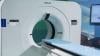
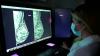
Yael Eshet, M.D., MSc, a diagnostic radiology specialist atSheba Medical Centerin Israel, was the lead author on a recent study that showedCOVID-19 (SARS-CoV-2)vaccine adenopathy can persist more than 6 weeks. This swelling of lymph nodes is similar to what is seen cancer and infections and the new findings show it can last longer than 7-10 weeks. The current recommended time people should delay medical imaging is 6 weeks after receiving a COVID vaccine to avoid a misdiagnosis,[2] but this new study shows there is increased inflammation shown on PET-CT imaging for much longer.

These were the findings in theRadiologypublished study"Prevalence of Increased FDG PET/CT Axillary Lymph Node Uptake Beyond 6 Weeks after mRNA COVID-19 Vaccination."[1]

Researchers using fluorodeoxyglucose (FDG)-positron emission tomography (PET)have found increased FDG uptake in the lymph nodes of patients 7-10 weeks past their second mRNA-based Pfizer-BioNTech COVID-19 vaccination. This new information indicates a persistent immune response that could be mistaken on imaging exams for serious conditions like lymphoma over a much longer period of time.

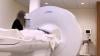
Recent recommendations for post-vaccine lymphadenopathy advise scheduling routine imaging, such as screening mammography, before, or at least 6 weeks after, the final vaccination dose to eliminate false positive results. However, this new research showed that avid axillary lymph node uptake was present beyond 6 weeks after the second vaccination in more than 29% of the patients in the study cohort.

The authors stated “This study shows that avid axillary lymph node uptake on FDG PET/CT can be detected in more than a quarter of our patient population even beyond 6 weeks after the second dose of the mRNA-based COVID-19 vaccination. Compared to a previous study showing normalization of FDG uptake within 40 days of receiving an inactivated H1N1 influenza vaccine, we found uptake persistence even at 70 days. Physicians should be aware of this potential pitfall.”

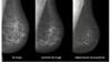
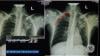
Some images in this video are from anotherRadiologystudy, which showed PET tracer uptake at the COVID vaccine injection site and other examples of axillary adenopathy.[3]

Related COVID Vaccine Axillary Adenapathy Content:

COVID-19 Vaccine Can Cause False Positive Cancer Diagnosis

Help Spread Awareness of Potential COVID-19 Vaccine Imaging Side-effects

视频:COVID Vaccine May Cause Enlarged Lymph Nodes on Mammograms— Interview with Constance "Connie" Lehman, M.D.

COVID-19 Vaccination Axillary Adenopathy Detected During Breast Imaging

PHOTO GALLERY: How COVID-19 Appears on Medical Imaging

CMS Now Requires COVID-19 Vaccinations for Healthcare Workers by January 4

Find more radiology related COVID content

References:

1.Yael Eshet, Noam Tau1, Yousef Alhoubani, Nayroz Kanana, Liran Domachevsky, Michal Eifer。mRNA COVID-19疫苗接种6周后FDG PET/CT腋窝淋巴结摄取增加的发生率放射学。Published Online:Apr 27 2021https://doi.org/10.1148/radiol.2021210886.

2.Constance D. Lehman, Leslie R. Lamb, and Helen Anne D'Alessandro. Mitigating the Impact of Coronavirus Disease (COVID-19) Vaccinations on Patients Undergoing Breast Imaging Examinations: A Pragmatic Approach American Journal of Roentgenology. 10.2214/AJR.21.25688.

3.可以Özütemiz, Luke A. Krystosek, An L. Church, Anil Chauhan, Jutta M. Ellermann, evdio Domingo-Musibay, Daniel Steinberger。COVID-19疫苗接种者的淋巴结病:肿瘤患者的诊断困境。放射学。Published Online:Feb 24 2021https://doi.org/10.1148/radiol.2021210275.

Point-of-Care Ultrasound (POCUS)| April 01, 2021

Here are two quick clinical examples of point-of-care ultrasound (POCUS) lung imaging and cardiac imaging using a GE Vscan Air device. The examples show an abnormal lung image with B-lines. The second clip shows a healthy heart in a parasternal color Doppler image.

The GE Healthcare Vscan Air is a cutting-edge, wireless pocket-sized ultrasound that provides crystal clear image quality, whole-body scanning capabilities, and intuitive software. The pocket-sized ultrasound system was originally introduced in 2010, and as of early 2021, there are over 30,000 Vscan systems in use. The new Vscan Air features a wireless ultrasound probe.

Read more in the articleGE Healthcare Unveils Vscan Air Wireless Handheld Ultrasound

Find more POCUS news and video

2022世界杯篮球预选赛赛程| March 26, 2021

Constance "Connie" Lehman, M.D.博士、乳房成像主任、马萨诸塞州总医院2022世界杯篮球预选赛赛程雅芳综合乳房评估中心联席主任、哈佛医学院放射学教授解释了问题,并为接种COVID-19疫苗并需要进行乳房x光检查的女性提出了指导方针。在疫苗发布后的头三个月里,已经有许多疫苗导致淋巴结肿大的病例报告。这通常会引发乳腺癌的危险信号,但对许多接受疫苗的女性来说是正常的,因为她们的身体免疫系统正在对抗病毒。

Lehman said cases reports of axillary adenopathy have been identified on breast imaging aftercoronavirus disease (COVID-19)vaccination and are rising. Lehman et al. proposed a pragmatic management approach in arecent article in theAmerican Journal of Roentgenology (AJR).[1]

In the settings of screening mammography, screening MRI and diagnostic imaging work-up of breast symptoms, with no imaging findings beyond unilateral axillary adenopathy ipsilateral to recent (prior six weeks) vaccination, they report the adenopathy as benign with no further imaging indicated if no nodes are palpable six weeks after the last vaccine dose.

For patients with palpable axillary adenopathy in the setting of ipsilateral recent vaccination, clinical follow-up of the axilla is recommended. In all these scenarios, axillary ultrasound is recommended if clinical concern persists six weeks after vaccination.

In patients with recent breast cancer diagnosis in the pre- or peri-treatment setting, prompt recommended imaging is encouraged as well as vaccination (in the thigh or contralateral arm). The recommendations align with the ACR BI-RADS Atlas and aim to: 1) reduce patient anxiety, provider burden, and costs of unnecessary evaluation of enlarged nodes in the setting of recent vaccination, and 2) avoid further delays in vaccinations and breast cancer screening during the pandemic.

Related Medical Imaging of COVID Content:

COVID-19 Vaccination Axillary Adenopathy Detected During Breast Imaging

CMS Now Requires COVID-19 Vaccinations for Healthcare Workers by January 4

PHOTO GALLERY: How COVID-19 Appears on Medical Imaging

视频:Imaging COVID-19 With Point-of-Care Ultrasound (POCUS)— Interview with Mike Stone, M.D.

视频:Use of Teleradiology During the COVID-19 Pandemic— Interview with John Kim, M.D.

Find more radiology related COVID content

Reference:

1.Constance D. Lehman, Leslie R. Lamb, and Helen Anne D'Alessandro. Mitigating the Impact of Coronavirus Disease (COVID-19) Vaccinations on Patients Undergoing Breast Imaging Examinations: A Pragmatic Approach American Journal of Roentgenology. 10.2214/AJR.21.25688

Magnetic Resonance Imaging (MRI)| March 19, 2021

Darryl B. Sneag, M.D., a radiologist and director of peripheral nerve MRI at theHospital for Special Surgery (HSS)in New York City, explains howartificial intelligence (AI)magnetic resonance imaging (MRI)reconstruction algorithms have cut imaging times by 50 percent. This has enabled his facility to maintain the same number of patients as it did prior to the pandemic, while still having time to sterilize the scanners after each patient.

Many radiology departments are now experiencing a backlog of cases due toCOVID-19shutdowns in 2020 and the limits on the number of patients that can be in the hospital for imaging exams due to pandemic containment precautions. Sneag said AI is now playing a role in helping streamline workflow.

HSS has 19 GE Healthcare MRI scanners and uses theAir Recon DL AI image reconstruction algorithm.这使得扫描时间更短,因此即使对核磁共振孔进行更深入的清理,每天也可以对与大流行前相同数量的患者进行成像。Sneag explains the algorithm has greatly helped with patient throughput, but the trade off is sometimes getting a ringing artifact on images.

HSS also uses GE'sAir Coil flexible pad MRI coils.这些线圈可以缠绕在患者周围,以提高舒适度,并使线圈更接近被成像的解剖结构。

Related MRI and COVID Content:

Business During COVID-19 and Beyond

Imaging Volumes Hold Steady Post COVID-19 Closures

GE Healthcare Addresses Growing Radiology Data Challenges at RSNA 2019

Technology is Driving the MRI Market

视频:How to Image COVID-19 and Radiological Presentations of the Virus

Top Trend Takeaways in Radiology From RSNA 2020

Post-COVID Pain or Weakness? Request an Ultrasound or MRI

Find more COVID radiology-related content

Coronavirus (COVID-19)| November 15, 2020

Margarita Revzin, M.D., MS, FSRU, FAIUM, associate professor of radiology and biomedical imaging, Yale University School of Medicine, abdominal and emergency imaging, radiologist, explains how different medical imaging modalities are used to image manifestations of theCOVID-19 (SARS-CoV-2)virus in patients. She is the lead author on a two-part article in the RSNA journalRadiographicsthat provides a comprehensive overview of coronavirus imaging.

The articles offer numerous case images from X-ray, ultrasound, computed tomography (CT), and magnetic resonance imaging (MRI). Revzin also discusses some of the radiology presentations and complications of the virus and which modalities can best image these features. Here are links to the two articles:

COVID-19的表现,第一部分:病毒发病机制和肺和血管系统并发症。

Multisystem Imaging Manifestations of COVID-19, Part 2: From Cardiac Complications to Pediatric Manifestations

虽然COVID-19主要影响呼吸系统,但也可能涉及其他器官。文章作者表示,影像学在本病所有表现及其相关并发症的诊断中起着至关重要的作用,正确利用和解释影像学检查是至关重要的。随着病毒的持续传播,对诊断性影像学特征、影像学特征、多系统累及以及影像学表现演变的全面了解对于有效的患者管理和治疗至关重要。在2020年秋季发表本系列文章之前,只有几篇文章全面描述了COVID-19的多系统影像学表现。作者提供了一个包容性的系统,系统的图像为基础的审查这种威胁生命和迅速传播的感染。在这篇文章的第1部分,作者讨论疾病的一般方面,重点是病毒学,病毒的病理生理学,和疾病的临床表现。第二部分重点介绍COVID-19的关键影像学特征,包括心脏、神经系统、腹部、皮肤、眼部和肌肉骨骼系统,以及儿童和妊娠相关的病毒表现。

Most of the images in the video are from the articles. Find more COVID medical imaging in thePHOTO GALLERY: How COVID-19 Appears on Medical Imaging

视频:Example of COVID Thrombosis on Ultrasound Imaging

Coronavirus (COVID-19)| February 09, 2021

Margarita Revzin, M.D., MS, FSRU, FAIUM, associate professor of radiology and biomedical imaging, Yale University School of Medicine, abdominal and emergency imaging, radiologist, explains how different medical imaging modalities are used to image manifestations of theCOVID-19 (SARS-CoV-2)virus in patients. She is the lead author on a two-part article in the RSNA journalRadiographicsthat provides a comprehensive overview of coronavirus imaging.

The articles offer numerous case images from X-ray, ultrasound, computed tomography (CT), and magnetic resonance imaging (MRI). Revzin also discusses some of the radiology presentations and complications of the virus and which modalities can best image these features. Here are links to the two articles:

COVID-19的表现,第一部分:病毒发病机制和肺和血管系统并发症。

Multisystem Imaging Manifestations of COVID-19, Part 2: From Cardiac Complications to Pediatric Manifestations

虽然COVID-19主要影响呼吸系统,但也可能涉及其他器官。文章作者表示,影像学在本病所有表现及其相关并发症的诊断中起着至关重要的作用,正确利用和解释影像学检查是至关重要的。随着病毒的持续传播,对诊断性影像学特征、影像学特征、多系统累及以及影像学表现演变的全面了解对于有效的患者管理和治疗至关重要。在2020年秋季发表本系列文章之前,只有几篇文章全面描述了COVID-19的多系统影像学表现。作者提供了一个包容性的系统,系统的图像为基础的审查这种威胁生命和迅速传播的感染。在这篇文章的第1部分,作者讨论疾病的一般方面,重点是病毒学,病毒的病理生理学,和疾病的临床表现。Part 2 focuses on key imaging features of COVID-19 that involve the cardiac, neurologic, abdominal, dermatologic and ocular, and musculoskeletal systems, as well as pediatric and pregnancy-related manifestations of the virus

Most of the images in the video are from the articles. Find more COVID medical imaging in thePHOTO GALLERY: How COVID-19 Appears on Medical Imaging

Related Medical Imaging of COVID Content:

视频:What Does COVID-19 Look Like in Lung CT Scans

PHOTO GALLERY: How COVID-19 Appears on Medical Imaging

视频:Imaging COVID-19 With Point-of-Care Ultrasound (POCUS)— Interview with Mike Stone, M.D.

视频:COVID Vaccine May Cause Enlarged Lymph Nodes on Mammograms— Interview with Constance "Connie" Lehman, M.D., Mass General Hospital

视频:Use of Teleradiology During the COVID-19 Pandemic— Interview with John Kim, M.D.

视频:Radiology Industry Responding to COVID-19— Interview with Jeffrey Bundy, Ph.D.

CT in a Box Helps Rapidly Boost Imaging Capability at COVID Surge Hospitals

视频:How China Leveraged Health IT to Combat COVID-19— Interview with Jilan Liu, M.D.

Find more radiology related COVID content

Coronavirus (COVID-19)| January 26, 2021

This is an example of aCOVID-19 (SARS-CoV-2)positive patient's lungcomputed tomography (CT)scan. The video scrolls through the image slices of the scan and shows the typical white, ground glass opacities (GGO) caused by COVID pneumonia. The pneumonia typically appears along the walls of each lobe of the lung, especially the chest wall and the lower portions of the lungs. This scan is from a Canon Aquilion Prime SP CT scanner and used Advanced intelligent Clear-IQ Engine (AiCE), an artificial intelligence-driven image reconstruction software to improve image quality of lower-dose scans. This was shown by Canon Medical as an exmaple of CT image quality for the virus at the 2020Radiological Society of North American (RSNA)meeting.

Read more about this system and its launch in 2020 to address COVID,Canon Medical Launches CT Solution for Patients with Viral Infectious Diseases.

视频:How to Image COVID-19 and Radiological Presentations of the Virusinterview with Margarita Revzin, M.D., associate professor of radiology and biomedical imaging, Yale School of Medicine.

Find more radiology clinical images of coronavirus in this photo gallery.

Find more radiology related COVID news and video

PET-CT| December 04, 2020

This is an example of Canon'sAdvanced intelligent Clear-IQ Engine (AiCE)AI-driven image reconstruction software that is now being used to improve image quality on the Canon Celesteion Prime PET/CT nuclear imaging system. Thedeep learningis used to enhance the iterative reconstruction used to reduce noise and sharped high contrast resolution onpositron emission tomography (PET)images from the digital PET detector used on the system.

This example is a whole-body FGD PET scan of a patient with a large BMI with lung cancer.

The Cartesion PrimePET/CTis the industry’s only air-cooled digital PET/CT, provides variable bed time (vBT) acquisition as a standard feature. This and the new FDA 510(k)-pending AiCE technology were highlighted at the 2020 Radiological Society of North America (RSNA) virtual meeting.

Find more RSNA news

Artificial Intelligence| December 02, 2020

Kirti Magudia, M.D.,Ph.D., an abdominal imaging and ultrasound fellow at the University of California San Francisco, explains how an automated deep learning analysis of abdominalcomputed tomography (CT)图像可以更精确地测量身体组成,并更好地预测主要心血管事件,如心脏病发作和中风,比整体体重或身体质量指数(BMI)更好。This was according to a study she presented at the 2020Radiological Society of North America (RSNA)virtual meeting.

与BMI(基于身高和体重)不同,腹部单轴CT切片可以显示皮下脂肪区、内脏脂肪区和骨骼肌区的体积。然而,手动测量这些单独的区域是费时且昂贵的。一个多学科研究团队,包括放射科医生、数据科学家和生物统计学家,开发了一种全自动人工智能(AI)方法,从腹部CT图像确定人体组成指标。

Statistical analysis demonstrated that visceral fat area was independently associated with future heart attack and stroke. BMI was not associated with heart attack or stroke.

Read more about this study

Find more RSNA news

沙特vs阿联酋比分预测| December 01, 2020

Treating cancer effectively often includes a combination of patient therapies. In recent years, technology advancements have led to a more efficient and personalized approach to treatment.Andrew Wilson, President of Oncology Informatics at Elekta, discussed the latest software advancements withITN

Remote Viewing Systems| November 28, 2020

Konica Minolta’s theme forRSNA 2020is Depth of Vision.ITNrecently talked withDavid Widmann, President and CEO ofKonica Minolta Healthcare Americas, about this focus and their key messages for customers andRSNAattendees.

X-Ray| November 28, 2020

Agfais looking to transformX-raywith new advancements in volumetric imaging, and with new mobile concepts and implementation ofintelligent toolsITNhad a conversation withGeorges Espadaon Transforming X-ray with Intelligent Tools.

Enterprise Imaging| November 23, 2020

Fujifilm's next generation secure server-side viewer platform extends acrossenterprise imagingareas to bring together radiology, mammography and cardiology into a single zero footprint platform.Bill Lacy, vice president of medical informatics forFujifilm Medical Systems USArecently talked withITNabout theirSynapse 7x platform

Coronavirus (COVID-19)| November 20, 2020

This video shows a computed tomography (CT) scroll through showing bowel ischemia and perforation (see arrows) due to superior mesenteric artery (SMA) thrombus inCOVID-19 (SARS-CoV-2)patient. Mesenteric artery thrombosis (MAT) is a condition involving occlusion of the arterial vascular supply of the intestinal system and is a severe and potentially fatal illness.

Superior mesenteric artery thrombosis (Red arrow) complicated by bowel ischemia and perforation in a 54-year-old man who presented to the emergency department with abdominal pain and was diagnosed with COVID-19. Contrast-enhanced CT images of the abdomen and pelvis show mucosal hyperenhancement involving the small bowel (green arrows).

Read more in the articleMultisystem Imaging Manifestations of COVID-19, Part 1: Viral Pathogenesis and Pulmonary and Vascular System Complications

Case example fromMargarita Revzin, M.D., associate professor of radiology and biomedical imaging, Yale School of Medicine.

视频:How to Image COVID-19 and Radiological Presentations of the Virus— Interview with Margarita Revzin, M.D.,

See more medical imaging of COVID-19 in the photo galleryHow COVID-19 Appears on Medical Imaging.

Related COVID Radiology Content:

视频:COVID-19 Pneumonia Chest CT Scan Scroll Through

CT Imaging of the 2019 Novel Coronavirus (2019-nCoV) Pneumonia

Chest CT Can Distinguish Negative From Positive Lab Results for COVID-19

视频:CT Sees Increased Use During COVID-19

Handheld Ultrasound Used to Monitor COVID-19 Patients With Cardiac Complications

Study Looks at CT Findings of COVID-19 Through Recovery

Using Lung X-rays to Diagnose COVID-19

视频:Use of Teleradiology During the COVID-19 Pandemic— Interview with John Kim, M.D.

Artificial Intelligence| November 11, 2020

Artificial Intelligence (AI)is becoming more common place in radiology practices, and emerging technologies are providing radiologists with sophisticated detection software to aid their reading and provide support for a busy workflow. With the progression of AI technology, vendors must look not only atwhatAI can do for the radiologist, buthowthe radiologist and the technician interact with that technology – the goal should be increasing accuracy while also positively improving workflow. GE Healthcare is working to improve radiology AI workflow in itsCentricity Universal Viewer.

Three key opinion leaders offers their views on what is needed to make AI more valauble and accessible to radiologists. These include:

Amy Patel, M.D.她是一位乳腺放射科医生,自由医院女性影像医学主任,密苏里大学堪萨斯城分校放射科助理教授。

Prof. Dr. Thomas Frauenfelder, M.D., vice chairman and professor of thoracic radiology, Institute for Diagnostic and Interventional Radiology, University of Zurich.

Randy Hicks, M.D., chief executive officer, Regional Medical Imaging.

Learn more about the Centricity Universal Viewer in the视频:How GE Healthcare’s Zero Footprint Remote Image Viewer Supports Clinical Care

Subscribe Now