News|Medical 3-D Printing| December 11, 2018

VA Puget Sound Health Care System and GE Healthcare are working to reduce the time it takes for radiologists to create 3-D-printed models and prosthetics from hours to minutes

GE Healthcare Partners With Department of Veterans Affairs to Accelerate 3-D Printing

December 11, 2018 — GE Healthcare and VA Puget Sound Health Care System, Washington, recently announced a partnership to accelerate the use ofmedical 3-D printingin healthcare. As part of their research agreement, GE Healthcare will provide software and workstations, and the VA will provide input on its use of the technology. Prior to this agreement, the VA has used 3-D software that is not designed for medical use. Now, the VA will use GE software designed specifically for the medical field – which is expected to reduce the time it takes to create 3-D models from hours to minutes.

VA Puget Sound provides comprehensive care to more than 110,000 veterans across its nine facilities in the Pacific Northwest. Building on its 3D printing network, VA Puget Sound and the Veterans Health Administration Innovators Network will integrate GE Healthcare’sadvanced visualizationAW VolumeShare workstations with 3-D printing software across its facilities in Seattle, San Francisco, Minneapolis, Cleveland and Salt Lake City. VA radiologists specializing in cardiology, oncology, orthopedics and other areas will use this technology and software to develop new 3-D imaging approaches and techniques to deliver improved precision healthcare for U.S. veterans.

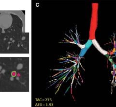
“The Veterans Health Administration has been on the forefront of bringing 3-D printing to the bedside, and we are thrilled to join forces with GE Healthcare to enhance and accelerate its adoption,” said Beth Ripley, M.D., Ph.D., VA Puget Sound radiologist, VA innovation specialist and VHA 3D Printing Advisory Committee chair. “For most radiologists, 3-D images are limited to reconstructions on a computer screen. By harnessing the power of 3-D printing with a rich data set, we are able to pull images out of the screen and into our hands, allowing us to interact with the data in a deeper way to fuel innovative, personalized care based on the unique needs of each of our patients.”

The use of 3-D medical printing in healthcare is still very much in its infancy, and software designed exclusively for the medical community is limited. Software designed to allow manual preparation of image data into 3-D printable files can be labor-intensive, requiring hours of work. Using GE Healthcare’s advanced visualization tool, specifically designed for the medical community, VA radiologists will be able to produce models of normal and pathological anatomy using automation techniques that will speed up the pre-3-D printing preparation work and the diagnostic process. This is expected to reduce the time it takes to create 3-D models from hours to minutes.

Three-dimensional printing is primarily used to manufacture orthopedic implants and guide surgical cutting, and peer-reviewed research on potential impact in patient care has expanded exponentially. Recent industry and regulatory advancements such as the establishment of clinical guidelines, 3-D printing reimbursement tracking codes, and the integration of technology and software are all expected to support the widespread adoption of point-of-care 3-D printing in hospitals.

For more information:www.gehealthcare.com

Related Content

The National Imaging Informatics Course-Radiology’s (NIIC-RAD) next term will resume online this fall from September 19 to September 23, 2022
News|Radiology Education

2022年8月9日-国家成像信息学课程-放射学(NIIC-RAD)下学期将在今年秋天恢复在线…

TimeAugust 09, 2022
arrow
Images show airway metrics. (A) Noncontrast-enhanced axial view chest CT image shows cross sections of segmental airways. (B) Inset shows segmented airway wall (green) and airway lumen (pink). (C) Segmented airway tree with color-coded branches. The resulting total airway count (TAC) and airway fractal dimension (AFD) are shown in C.
News|Lung Imaging

August 3, 2022 — Structural differences in lung airways between men and women may be the cause of differences in chronic ...

TimeAugust 03, 2022
arrow
The CARESTREAM Lux 35 Detector, a lightweight, glass-free wireless detector ergonomically designed with the comfort of patients and radiographers in mind, earned the 2022 Frost & Sullivan Global New Product Innovation Award
News|Digital Radiography (DR)

July 29, 2022 — Carestream Health’s focus on delivering innovative medical imaging solutions that address customer needs ...

TimeJuly 29, 2022
arrow
The benefits of adding whole breast automated ultrasound (ABUS) screening to clinical practice, enhancing cancer detection in the setting of dense breast tissue
Feature|2022世界杯篮球预选赛赛程| By Christine Book

In a detailed overview held during the Radiological Society of North America’s (RSNA) 2021 annual meeting, promising ...

TimeJuly 08, 2022
arrow
Here are excerpts from an in-depth, one hour discussion with a stellar panel of radiologists and biomedical imaging experts on the present, and future, state of artificial intelligence (AI) in radiology
Feature|Artificial Intelligence| By Christine Book

Among the hundreds of educational sessions and interesting exhibits at the Radiological Society of North America’s (RSNA ...

TimeJuly 07, 2022
arrow
Siemens Healthineers has announced the Food and Drug Administration (FDA) clearance of the Magnetom Free.Star
News|Magnetic Resonance Imaging (MRI)

July 5, 2022 — Siemens Healthineers has announced the Food and Drug Administration (FDA) clearance of the Magnetom Free ...

TimeJuly 05, 2022
arrow
The Radiological Society of North America (RSNA) reports that Coronary Artery Calcium (CAC) scoring with use of noncontrast electrocardiographically gated Computed Tomography (CT) is a fast, inexpensive and reproducible examination that effectively-predicts risk for development of cardiovascular diseases and guides preventive therapy in asymptomatic individuals
News|Cardiac Imaging

June 24, 2022 — The Radiological Society of North America (RSNA) reports that Coronary Artery Calcium (CAC) scoring with ...

TimeJune 24, 2022
arrow
Gradient Health, a medical technology company based in North Carolina, announced a new initiative which will give academic researchers open access to their vast and diverse medical imaging datasets
News|Artificial Intelligence

June 22, 2022 — Gradient Health, a medical technology company based in North Carolina, announced a new initiative which ...

TimeJune 22, 2022
arrow
After less than two years of data collection and processing, the Radiological Society of North America (RSNA) has successfully delivered over 30,000 de-identified imaging exams to the Medical Imaging and Data Resource Center (MIDRC), an open-access platform which publishes data to be used for research.
News|Radiology Imaging

June 21, 2022 — After less than two years of data collection and processing, the Radiological Society of North America ...

TimeJune 21, 2022
arrow
Flow diagram for feature selection and deep-radiomics model development. LASSO = least absolute shrinkage and selection operator, MLP = multilayer perceptron, Model-C = model with clinical features, Model-T = model with texture features, Model-TC = model with texture and clinical features, Model-D = model with deep features, Model-DC = model with deep and clinical features, Model-DT = model with deep and texture features, Model-DTC = model with deep, clinical, and texture features.
News|Artificial Intelligence

May 25, 2022 — A new method that combines imaging information with artificial intelligence (AI) can diagnose ...

TimeMay 25, 2022
arrow
Subscribe Now