Technology|Medical 3-D Printing| June 18, 2018

3D printed anatomical models can provide medical professionals enhanced visualization and can also be used for patient education

3D Systems Announces On Demand Anatomical Modeling Service

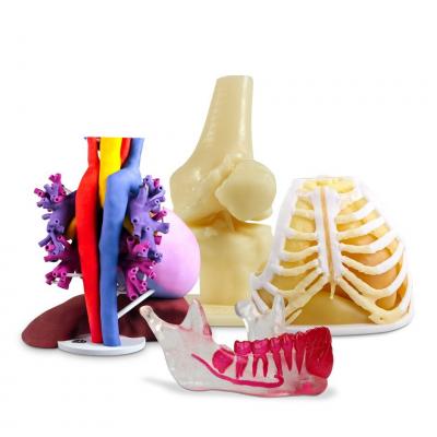
June 18, 2018 — 3D Systems announced availability of its new On Demand Anatomical Modeling Service. This new service provides a wide range of medical professionals with access to anatomical models3-D-printedfrom their 3-D digital files, enabling enhancedthree-dimensional visualizationfor surgical planning as well as patient education.

Obtaining a model requires only a few simple steps:

  • Medical professionals can easily upload a 3-D model file (i.e., STL, OBJ or PLY) to the company's On Demand Anatomical Models website. Customers can prepare model files with 3D Systems' D2P software or any commercially available software;
  • Customers select from a variety of materials from which to print depending on the use and desired areas to highlight in the model, and then request an instant quote; and
  • After reviewing the quote, the requestor can place the order in just one click, and the finished model will arrive in approximately five business days.

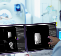
To further expedite the process, 3D Systems created a seamless connection between its D2P software and the On Demand Anatomical Models website. This end-to-end medical solution allows surgeons, radiologists, lab technicians and other medical professionals to quickly create accurate, digital 3-D anatomical models from medical imaging data.

D2P now includes a unique module for Volume VR, enabling the upload and launch of the entire patient scan into a 3D Virtual Reality environment without any preprocessing of the data. This development allows the user to walk through their scans and see an enhanced view of their patient's anatomy, control layer visualization and cut cross sections in any direction. Further enhancements include improved mesh creation options, import and alignment of external mesh file into patient scan, and 3-D PDF generation.

In addition to this new service, 3D Systems continues to offer full-service virtual surgical planning and anatomical modeling services. For medical professionals wishing to produce models for diagnostic purposes and pre-surgical planning, the company provides a Patient-Specific Anatomical Modeling option. To utilize 3D Systems' anatomical modeling service, a medical professional provides a computed tomography (CT) or magnetic resonance imaging (MRI) scan of their patient to the company's team of experts at its Healthcare Technology Center in Littleton, Colo. In turn, the company's biomedical engineers process the data, design the model and 3-D-print it at the facility. The finished model is then shipped to the medical professional for use in pre-surgical planning, pre-surgical rehearsal and for educational purposes. Certain materials can also be used in a sterile environment such as an operating room for consultation during a procedure.

For more information:www.3dsystems.com

Related Content

Digital surgery pioneer conducts 40-patient study with ActivSight Intraoperative Imaging Module to demonstrate advanced visualization use cases in the operating room
News|Virtual and Augmented Reality

May 24, 2022 — Activ Surgical, a digital surgery pioneer, today announced the successful completion of an Institutional ...

TimeMay 24, 2022
arrow
Adaptiiv is pioneering new approaches to bring personalization at scale to patients all over the world and is collaborating with HP and Varian to advance these efforts and meet evolving customer needs
News|Medical 3-D Printing

May 11, 2022 — Adaptiiv Medical Technologies is collaborating with HP Inc. and Varian, a Siemens Healthineers company ...

TimeMay 11, 2022
arrow
Videos|Radiation Oncology

Douglas E. Holt, M.D., a radiation oncologist at Eastern Idaho Regional Medical Center, explains the use of 3-D virtual ...

TimeFebruary 02, 2022
arrow
Videos|Computed Tomography (CT)

Cynthia McCollough, Ph.D., director of Mayo Clinic's CT Clinical Innovation Center, explains how photon-counting ...

TimeJanuary 27, 2022
arrow
Cydar EV Maps有助于血管内手术的规划、实时指导和术后回顾。
News|Mobile C-Arms

January 18, 2022 – Philips Healthcare announced physicians will now have access to advanced new 3D image guidance ...

TimeJanuary 18, 2022
arrow
在这两种情况下,外科医生都使用了飞利浦集成的Spine Suite解决方案,该解决方案为该公司的Azurion混合手术室(OR)提供了ClarifEye,这是业界首创的解决方案,将低x射线剂量下的2D和3D可视化与3D AR导航结合到一个系统中。
News|Virtual and Augmented Reality

December 8, 2021 — Royal Philips, a leader in health technology, announced that the first patients have been ...

TimeDecember 08, 2021
arrow
Realize Medical, creators of Elucis, an all-in-one virtual reality platform for medical modeling, visualization and remote collaboration, announces their first in-person exhibit at RSNA in Chicago
News|Virtual and Augmented Reality

2021年11月15日—医学建模一体化虚拟现实平台Elucis的创造者Realize Medical…

TimeNovember 15, 2021
arrow
Canon's Vitrea PACS enterprise imaging system was one of several systems demonstrated at HIMSS 2021 that had easily modified hanging protocols. This included ease of use to customize what each radiologists prefers, including slice thickness. #HIMSS #HIMSS21
Feature|Enterprise Imaging

Taking advantage of new technology advances, several radiology PACS, enterprise imaging and cardiovascular information ...

TimeNovember 04, 2021
arrow
The Siemens Naeotom Alpha is the first commercialized photon-counting CT scanner. It gained FDA clearance Sept. 30, 2021. The image shows the system undergoing phantom testing with the outer covers off, showing the motion of the X-ray source and detector ring. Image by Deutscher Zukunftpreis/Ansgar Pudenz.
Feature|Computed Tomography (CT)| By Dave Fornell, ITN Editor

The U.S. Food and Drug Administration (FDA) cleared the world's first photon-counting computed tomography (CT) scanner ...

TimeNovember 04, 2021
arrow
Cardiac CTA derived left atrium emptying fraction (LAEF) improves predictive performance of established clinical risk scores and may be used to assess TAVR patient risk during pre-procedure workup and post-procedural surveillance. These images show LAVmax (A and B), LAVmin (C and D), and LAEF using cardiac CTA. LAEF was calculated as (LAVmax-LAVmin)/LAVmax x 100. Given LAVmax of 109 mL and LAVmin of 80 mL, the LAEF was calculated to be 27%. Image courtesy of AJR.
News|Computed Tomography (CT)

October 14, 2021 — Cardiac computed tomography angiography (CTA) derived left atrium emptying fraction (LAEF) improves ...

TimeOctober 14, 2021
arrow
Subscribe Now