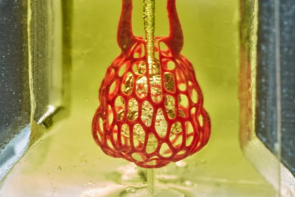
Bioprinting research from the lab of Rice University bioengineer Jordan Miller featured a proof-of-principle — a scale-model of a lung-mimicking air sac with airways and blood vessels that never touch yet still provide oxygen to red blood cells. Image courtesy of Jordan Miller/Rice University.
May 3, 2019 — Bioengineers have cleared a major hurdle on the path to3-D printing用生物打印组织的突破性技术来替代器官。这项新发明使科学家们能够创造出精巧的纠缠的血管网络,模仿人体的血液、空气、淋巴和其他重要液体的自然通道。
The research is featured on the cover ofScience.1 It includes a visual proof-of-principle — a hydrogel model of a lung-mimicking air sac in which airways deliver oxygen to surrounding blood vessels. Also reported are experiments to implant bioprinted constructs containing liver cells into mice.
The work was led by bioengineersJordan Millerof Rice University andKelly Stevensof the University of Washington (UW). It included 15 collaborators from Rice, UW, Duke University, Rowan University and Nervous System, a design firm in Somerville, Mass.
"One of the biggest road blocks to generating functional tissue replacements has been our inability to print the complex vasculature that can supply nutrients to densely populated tissues," said Miller, assistant professor of bioengineering at Rice'sBrown School of Engineering. "Further, our organs actually contain independent vascular networks — like the airways and blood vessels of the lung or the bile ducts and blood vessels in the liver. These interpenetrating networks are physically and biochemically entangled, and the architecture itself is intimately related to tissue function. Ours is the first bioprinting technology that addresses the challenge of multivascularization in a direct and comprehensive way."
Stevens, assistant professor of bioengineering in the UW College of Engineering, assistant professor of pathology in the UW School of Medicine, and an investigator at theUW MedicineInstitute for Stem Cell and Regenerative Medicine, said multivascularization is important because form and function often go hand in hand.
史蒂文斯说:“组织工程已经为此挣扎了一代人。”“有了这项工作,我们现在可以更好地问,‘如果我们能打印出看起来和呼吸更像我们身体健康组织的组织,那么它们在功能上是否也会更像那些组织?’这是一个重要的问题,因为生物打印组织的功能将影响到它在治疗上的成功程度。"
The goal ofbioprintinghealthy, functional organs is driven by the need for organ transplants. More than 100,000 people are on transplant waiting lists in the United States alone, and those who do eventually receive donor organs still face a lifetime of immune-suppressing drugs to prevent organ rejection. Bioprinting has attracted intense interest over the past decade because it could theoretically address both problems by allowing doctors to print replacement organs from a patient's own cells. A ready supply of functional organs could one day be deployed to治疗全球数百万患者。
"We envision bioprinting becoming a major component of medicine within the next two decades," Miller said.
史蒂文斯说:“肝脏特别有趣,因为它能执行令人难以置信的500种功能,可能仅次于大脑。”“肝脏的复杂性意味着,目前没有任何机器或疗法可以在肝脏功能衰竭时替代它的所有功能。生物打印的人体器官可能有一天会提供这种治疗。"
To address this challenge, the team created a new open-source bioprinting technology dubbed the "stereolithography apparatus for tissue engineering" (SLATE). The system uses additive manufacturing to make soft hydrogels one layer at a time.
Layers are printed from a liquid pre-hydrogel solution that becomes a solid when exposed to blue light. Adigital light processingprojector shines light from below, displaying sequential 2-D slices of the structure at high resolution, with pixel sizes ranging from 10-50 microns. With each layer solidified in turn, an overhead arm raises the growing 3-D gel just enough to expose liquid to the next image from the projector. The key insight by Miller and Bagrat Grigoryan, a Rice graduate student and lead co-author of the study, was the addition of food dyes that absorb blue light. These photoabsorbers confine the solidification to a very fine layer. In this way, the system can produce soft, water-based, biocompatible gels with intricate internal architecture in a matter of minutes.
Tests of the lung-mimicking structure showed that the tissues were sturdy enough to avoid bursting during blood flow and pulsatile "breathing," a rhythmic intake and outflow of air that simulated the pressures and frequencies of human breathing. Tests found that red blood cells could take up oxygen as they flowed through a network of blood vessels surrounding the "breathing" air sac. This movement of oxygen is similar to the gas exchange that occurs in the lung's alveolar air sacs.
To design the study's most complicated lung-mimicking structure, which is featured on the cover of Science, Miller collaborated with study co-authors Jessica Rosenkrantz and Jesse Louis-Rosenberg, co-founders of Nervous System.
In the tests of therapeutic implants for liver disease, the team 3-D-printed tissues, loaded them with primary liver cells and implanted them into mice. The tissues had separate compartments for blood vessels and liver cells and were implanted in mice with chronic liver injury. Tests showed that the liver cells survived the implantation.
Miller said the new bioprinting system can also produce intravascular features, like bicuspid valves that allow fluid to flow in only one direction. In humans, intravascular valves are found in the heart, leg veins and complementary networks like the lymphatic system that have no pump to drive flow.
"With the addition of multivascular and intravascular structure, we're introducing an extensive set of design freedoms for engineering living tissue," Miller said. "We now have the freedom to build many of the intricate structures found in the body."
Miller and Grigoryan are commercializing key aspects of the research through a Houston-based startup company called Volumetric. The company, which Grigoryan has joined full time, is designing and manufacturing bioprinters and bioinks.
Miller, a longstanding champion of open-source 3-D printing, said all source data from the experiments in the publishedSciencestudy are freely available. In addition, all 3-D printable files needed to build the stereolithography printing apparatus are available, as are the design files for printing each of the hydrogels used in the study.
"Making the hydrogel design files available will allow others to explore our efforts here, even if they utilize some future 3-D printing technology that doesn't exist today," Miller said.
Miller said his lab is already using the new design and bioprinting techniques to explore even more complex structures.
"We are only at the beginning of our exploration of the architectures found in the human body," he said. "We still have so much more to learn."
For more information:www.sciencemag.org
Related 3-D Printing Content
The Future of 3-D Printing in Medicine
Reference
 May 11, 2022