News|Lung Imaging| July 13, 2022

Initial results from clinical trial on lung lesion detection will be presented at ECR 2022

KA Imaging, a company that develops innovative X-ray imaging solutions, announced the initial results from a study examining the diagnostic value of single-exposure dual-energy subtraction radiography in lung lesion detection

July 13, 2022 —KA Imaging, a company that develops innovativeX-rayimaging solutions, announced the initial results from a study examining the diagnostic value of single-exposure dual-energy subtractionradiographyinlung lesiondetection.

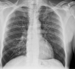
In the study, a radiologist blinded to standard-of-careCTresults was first asked to identify anomalies in the conventional X-ray. The same task was given right after that, with supplemented single-exposure dual-energy images. Quoting directly from the poster, “lesion visibility reportedly increased in 45% of the cases when supplemental dual-energy images were included1.” Findings were validated using CT.

“Lung cancer is the leading cause of cancer death; unfortunately, statistics show that it is rarely detected early,” explainsKarim S. Karim, CTO of KA Imaging. “The promising results from this trial show that spectral images can play an important role in earlier detection for better outcomes,” saysAmol Karnick, President and CEO.

The study will be presented in an e-poster at this year’s European Congress of Radiology (ECR). In addition to the e-poster at the scientific congress, KA Imaging is part of the technical exhibition at Expo X4, booth 406.

ECR is one of the largest medical meetings in Europe and the second-largest radiological meeting in the world. ECR attendees span all areas of the radiology arena including radiology professionals, radiographers, physicists, industry representatives, and press reporters for both the medical and consumer press.

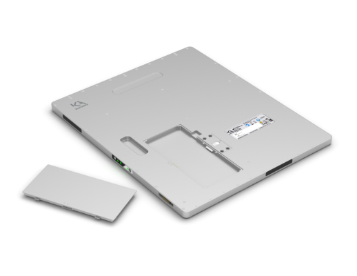
Recently, KA Imaging announced that its patented single-exposure dual-energy technology is now branded SpectralDR. The SpectralDR technology enables dual-energy subtraction, providing bone and tissue differentiation with a single standard X-ray exposure. It acquires three images simultaneously (DR, bone and soft tissue dual-energy X-ray images). The technology mimics the workflow, dose and techniques of state-of-the-art mobile DR X-ray detectors.

For more information:www.kaimaging.com

Find more ECR22 coverage here

Related Content

The U.S. Food and Drug Administration approved Enhertu (fam-trastuzumab-deruxtecan-nxki), an IV infusion for the treatment of patients with unresectable (unable to be removed) or metastatic (spread to other parts of the body) HER2-low breast cancer.
News|2022世界杯篮球预选赛赛程

August 8, 2022 — The U.S. Food and Drug Administration approved Enhertu (fam-trastuzumab-deruxtecan-nxki), an IV ...

TimeAugust 08, 2022
arrow
Dr. Milena Cavic, Institute for Oncology and Radiology of Serbia
News|Lung Imaging

August 8, 2022 — Representatives from the Diagnostics Working Group of the IASLC Early Detection and Screening Committee ...

TimeAugust 08, 2022
arrow
David Gerber, MD
News|Lung Imaging

August 8, 2022 — A clinical trial is only as powerful as its participants. For years, researchers have struggled to fill ...

TimeAugust 08, 2022
arrow
Chest with AI Brightness and Contrast (AIBC), using Helix 2.2 Advanced Imaging Processing.
News|X-Ray

August 5, 2022 — GE Healthcare released its most advanced fixed X-ray system yet, the next-generation Definium 656 HD ...

TimeAugust 05, 2022
arrow
Xoran Technologies announces that last month it received FDA 510(k) clearance for TRON — a truly mobile, full-body fluoroscopy, computed tomography (CT) X-ray system.
News|Computed Tomography (CT)

August 4, 2022 — Xoran Technologies announces that last month it received FDA 510(k) clearance for TRON — a truly mobile ...

TimeAugust 04, 2022
arrow
EHR screenshot showing sidebar text displayed to referring clinicians after placing orders for body CT (neck, chest, or abdomen and pelvis), describing iohexol shortage and providing appropriate strategies for iodinated contrast media conservation.
News|Contrast Media

August 3, 2022 — According to an open-access article in ARRS’ American Journal of Roentgenology (AJR), electronic health ...

TimeAugust 03, 2022
arrow
Images show airway metrics. (A) Noncontrast-enhanced axial view chest CT image shows cross sections of segmental airways. (B) Inset shows segmented airway wall (green) and airway lumen (pink). (C) Segmented airway tree with color-coded branches. The resulting total airway count (TAC) and airway fractal dimension (AFD) are shown in C.
News|Lung Imaging

August 3, 2022 — Structural differences in lung airways between men and women may be the cause of differences in chronic ...

TimeAugust 03, 2022
arrow
Bracco诊断公司宣布,美国食品和药物管理局(FDA)授予Iomeron®(iomeprol注射液)进口自由裁量权,以解决目前碘化造影剂短缺的问题。
News|Contrast Media

August 2, 2022 – A new update has been announced as the radiology world continues to address supply disruptions of ...

TimeAugust 02, 2022
arrow
The American Lung Association created LUNG FORCE, a national movement to defeat lung cancer, the leading cancer killer of women and men.
Feature|Lung Imaging

August 1, 2022 — In recognition of World Lung Cancer Day, the American Lung Association announced new patient resources ...

TimeAugust 01, 2022
arrow
UNC School of Medicine researchers documented significant decrease in neural flexibility across the entire brain of children with ADHD and throughout sub-networks considered crucial to cognitive flexibility
News|Magnetic Resonance Imaging (MRI)

July 27, 2022 — Multitasking is not just an office skill. It’s key to functioning as a human, and it involves something ...

TimeJuly 27, 2022
arrow
Subscribe Now