
BLOG: Assessing Anatomy in Motion with Dynamic Digital Radiography

DDR images provide a dynamic look at anatomical structures as they move.
A standard X-ray provides tremendous value to a clinician for its ability to quickly provide a static image of anatomical structures. There are limitations, however, to static exams. Multiple conventional X-rays are often needed to show a joint in flexed and extended positions. A single static image cannot provide the details of how a joint moves between flexion and extension points or tell the complete story.
Dynamic digital radiography(DDR) adds dynamic, or motion, capability to X-ray technology that captures sequential radiographic images in a single exam, enabling clinicians to observe the dynamic interaction of anatomical structures in motion over time. Unlike fluoroscopy, X-ray with DDR is not viewed in real-time, so an exam can be performed by a radiologic technologist without a physician present in the exam room.
DDR is an enhanced version of a standard digital radiography system that acquires up to 15 frames per second for as long as 20 seconds, resulting in a maximum of 300 X-ray images with a dose equivalent to about two standard X-rays. With DDR, radiation is lower than fluoroscopy or CT, and requires a shorter exam time than MRI.
Applications in Pulmonology
The benefits of DDR are being explored across a variety of disciplines. In pulmonology, DDR can be used to visualize and quantify lung function in relationship to surrounding structures. “You can watch the patient’s muscles as they breathe – as they inhale and as they exhale,” saidAlex Kagen, MD,site chair and associate professor of diagnostic, molecular and interventional radiology atMount Sinai MorningsideandMount Sinai West in New York City. “You can watch how much air is flowing in and flowing out.”
Dynamic Digital Radiography opens up potential forartificial intelligenceand various analytic applications to process the exam, quantify movement and track changes over time. “It’s taking the simple chest x-ray and pairing it with high end technology to now produce a functional exam that wasn’t present previously,” Kagen said.
Applications in Orthopedics
In orthopedics, DDR can be used to assess instability, musculoskeletal injury, sources of pain, and treatment follow-up of any joint throughout its range of motion – whether it be the neck, spine, shoulders or knees. It can also be a helpful tool for postoperative evaluation of movement in place of a more expensive CT or MRI exam.
“The fact that you’re seeing how the joint itself is moving when a patient is having pain is an inherent advantage,” saidEric Wagner, MD, MSc, assistant professor and director of upper extremity research atEmory Healthcare. “Most of the time, the patient is not coming to you because their shoulder hurts at rest; they’re coming to you because their shoulder hurts when they’re trying to move it for certain activities. This imaging allows you to see how the joint is moving during these activities.”
For Wagner, the difference between traditional X-ray and DDR is like learning a procedure from a book versus watching a video tutorial. “It’s so much more helpful, so much more useful when I'm trying to decide what is going on with that patient and how best to treat them,” Wagner said. “I have little doubt that this disruptive technology will eventually change the way we evaluate and manage musculoskeletal injuries.”
The Clinical Value of DDR
DDR的使用也被探索作为其他应用的有效工具,包括吞咽研究用于语言治疗,或作为在嗅测中评估膈肌运动的透视检查的替代品。
柯尼卡美能达医疗保健公司的数字放射摄影主任Guillermo Sander说:“临床医生现在可以通过动态x射线看到更多。”“由于DDR是使用相同x射线系统的另一种x射线技术,设备和技术人员的工作流程是相同的。它只是增加了一分钟的时间。”DDR可用于执行传统x射线检查的数字x光摄影系统,为医院和实践提供了一种具有成本效益的解决方案。
“For many physicians, DDR provides a more complete diagnosis and has become a key differentiator for their practice,” Sander said. “It introduces a technology with clinical value that also has the benefit of a CPT code for reimbursement.”
In the hospital environment, DDR adds value to what can be done. For patients, there is a wow factor when they see the motion in their X-ray. Sander says that anecdotal feedback suggests that patients who see a dynamic X-ray are more likely to understand their condition and adhere to their rehabilitation plan.
“Adding movement gives new insight, which makes it easier for clinicians to adopt technology and make it useful,” Sander said. “There is clinical value, economic value, technological value and patient appeal. This is a technology worth exploring.”
Editor’s note:This blog is the first in athree-part seriesabout innovative advancements in radiology.
Related DR content:
Guiding Diagnosis and Treatment of Musculoskeletal Conditions With Dynamic X-ray
Related Content

August 11, 2022 — Bracco Imaging S.p.A. has announced the renewal of the “Bracco Fellowships” initiative, in partnership ...
 August 11, 2022
August 10, 2022 — Mindray North America, a global leader and developer of healthcare technologies and solutions for ...
August 10, 2022
August 5, 2022 — GE Healthcare released its most advanced fixed X-ray system yet, the next-generation Definium 656 HD ...
August 05, 2022
August 4, 2022 — Xoran Technologies announces that last month it received FDA 510(k) clearance for TRON — a truly mobile ...
August 04, 2022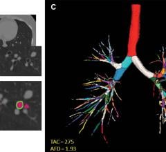
August 3, 2022 — Structural differences in lung airways between men and women may be the cause of differences in chronic ...
August 03, 2022
August 1, 2022 — In recognition of World Lung Cancer Day, the American Lung Association announced new patient resources ...
August 01, 2022
Here is what you and your colleagues found to be most interesting in the field of medical imaging during the month of ...
August 01, 2022
July 29, 2022 — Carestream Health’s focus on delivering innovative medical imaging solutions that address customer needs ...
July 29, 2022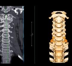
July 29, 2022 — The Radiological Society of North America (RSNA), in collaboration with the American Society of ...
July 29, 2022