
Dave Fornell is the editor of Diagnostic & Interventional Cardiology magazine and assistant editor for Imaging Technology News magazine.
Trends in Cardiovascular CT Imaging
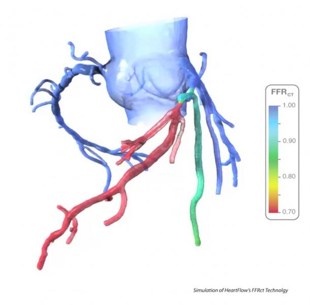
An example of FFR-CT, which uses supercomputing algorithms to determine a virtual assessment of hemodynamic flow through all coronary artery segments.
Here is a recap of some of the top trends and new technology at the Society of Cardiovascular Computed Tomography (SCCT) 2015 annual meeting in Las Vegas.
Watch video Editor’s Choice of the Most Innovative News Technology at SCCT 2015
Watch video on key trends from the SCCT President
Last November, the U.S. Food and Drug Administration (FDA) cleared the use of fractional flow reserve-CT (FFR-CT) software used to noninvasively virtually assess the blood flow through plaque narrowings of coronary vessels to see if the lesions have any hemodynamic significance. This may eliminate the need to perform invasive FFR readings in the cath lab, since the FFR-CT has a close correlation with the catheter-based technology. Catheter FFR is currently the gold standard for assessing the functional significance of lesions and to justify the need for stents. FFR-CT is viewed by many luminaries at SCCT as a potential paradigm shift in how chest pain and cardiac patients will be assessed in the future. This technology was definitely the poster child for SCCT 2015, and the subject of numerous packed sessions.
Watch a video interview on FFR-CT
CT perfusion imagingwas also a big topic of discussion, with numerous clinical trials now showing it can offer a more accurate functional assessment of blood flow in the heart without the need for a nuclear exam, stress testing or MRI.
新的临床试验数据继续表明,通过CT检查进行冠状动脉钙化评分的患者风险分层的准确性。钙评分评估似乎重新引起了人们的兴趣,它可以被用于类似于常规乳房x光检查来监测心血管疾病风险。数据显示,CT显示冠状动脉无钙,表明患者至少7年健康状况良好。定期的钙扫描可以用来确定患者是否需要开他汀类药物,并监测无症状的有冠状动脉疾病家族史的患者。SCCT还在开发一种类似于用于乳房的BI-RADS或用于肺癌评估的BI-RADS的放射学RADS风险评分系统,以促进未来这种类型的筛查。
Watch a video explaining how calcium scoring may add value to healthcare
Cardiac CT has become the gold standard imaging modality for assessing patients for transcatheter aortic valve replacement (TAVR). The CT images also are used for planning treatment, valve sizing, and both multiplanar and 3-D CT images are used to help guide the procedures. TAVR and CT’s roll with increasingly more complex transcatheter structural heart interventions was a major topic of discussion in sessions and on the expo floor. Edward’s Life Sciences, maker of the Sapien TAVR device, exhibited for the first time this CT specialty meeting, driving home the fact that these devices and imaging technologies go hand-in-hand.
Other big topics of discussion included the use of CT as a first line assessment to rule out coronary disease with chest pain patients in the emergency department, the utility of spectral CT imaging and 3-D printing.
Video on the use of CT to assess ED chest pain patients
Video on how 3-D cardiac printing is used at Henry Ford Hospital
Related Content

2022年8月8日- IASLC早期发现和筛查委员会诊断工作组的代表……
 August 08, 2022
August 4, 2022 — Xoran Technologies announces that last month it received FDA 510(k) clearance for TRON — a truly mobile ...
August 04, 2022
August 3, 2022 — According to an open-access article in ARRS’ American Journal of Roentgenology (AJR), electronic health ...
August 03, 2022
August 2, 2022 – A new update has been announced as the radiology world continues to address supply disruptions of ...
August 02, 2022
July 29, 2022 — Siemens Healthineers has announced the Food and Drug Administration (FDA) clearance of the ARTIS icono ...
July 29, 2022
July 25, 2022 — Dunlee unveiled its new oncology bundles for the first time this year, launched at the 2022 European ...
July 25, 2022
July 18, 2022 — 3DR Labs, an Accumen company, today announced the launch of its Cardiac Center of Excellence and Imaging ...
July 19, 2022
July 18, 2022 — Fresenius Kabi announced today it will introduce a portfolio of generic contrast media agents in the ...
July 18, 2022
July 13, 2022 — KA Imaging, a company that develops innovative X-ray imaging solutions, announced the initial results ...
July 13, 2022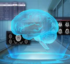
July 12, 2022 — The DeepCT is a leading AI-driven system developed by Deep01 to identify abnormal CT scan images ...
July 12, 2022