March 23, 2018 — A relatively inexpensive3-D-printed modelof a patient's blood vessels is as effective as current commercially available models for training medical students ininterventional radiologyvascular access, according to astudypresented at the Society of Interventional Radiology's 2018 Annual Scientific Meeting, March 17-22 in Los Angeles.
"We've come up with a viable method for creating something that's inexpensive and also customizable to individual patients," said Alexander Sheu, M.D., an interventional and diagnostic radiology resident at Stanford University School of Medicine, and lead author of the study. "The current model used to train medical students lacks the ability to replicate a patient's anatomy. Our 3-D-printed model will provide students a morerealistic experience, allowing for better preparation before they perform procedures on real patients."
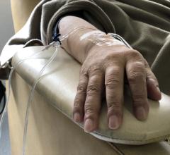
Interventional radiologists commonly treat patients using less-invasive options to surgery that involve inserting a catheter through a major artery under ultrasound guidance in order to reach internal organs or blood vessels. The researchers tested medical students' comfort in using a 3-D-printed model, compared to commercially available models, to simulate ultrasound-guided access through the femoral artery in the groin.
Thirty-two students were randomized to practice with the 3-D-printed model or the commercial model in a simulation experience developed by the authors of the study. Prior to the simulation exercise, 73 percent of the 3-D group and 76 percent of the commercial-model group indicated that they did not feel confident in performing the procedure. After the training, most of the 3-D model and commercial model trainees agreed that their respective models were easy to use (93.3 percent and 94.1 percent) and helpful for practice (93.3 percent and 94.1 percent). Additionally, confidence in performing the procedure, known as femoral artery access, increased a similar amount in both groups.
"Now that we know that a 3-D-printed model is just as effective at training medical students in this type of procedure, this simulation experience can be made available to even more trainees and potentially improve procedural skills for residents, fellows and attendees," said Sheu. "We foresee this really making an impact in the world of interventional radiology training."
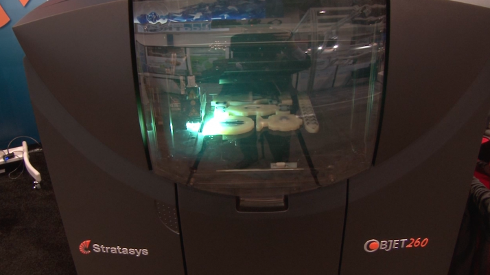
Medical simulation exercises are playing an increasingly larger role in medical training; especially in the field of interventional radiology. Many commercially available devices cost between $2,000 and $3,000 each, while 3-D printing has the ability to produce practice models inexpensively and more realistically, the authors said.
The 3-D printing technology can reproduce a patient's exact vessels based on a computed tomography (CT) scan and produce an ultrasound-compatible vascular access model that is unique to that patient's anatomy. To adapt the 3-D printing technology to their needs, the researchers used a tissue-mimicking material that was durable to withstand punctures, but still felt realistic. This tailoring allows trainees to practice with variations in anatomy before they encounter them during a procedure, which may help to lower complication rates, researchers said.
As a result of these findings, the research team aims to extend this training to resident and fellow trainees and to study additional possible benefits of these devices. In addition, the team may develop 3-D-printed models for other parts of the body with arteries accessed in interventional radiology.
For more information:www.sirmeeting.org
 August 08, 2022