December 1, 2021 — In the first MRI-based study to investigate prenatal alcohol exposure, researchers found significant changes in the brain structure of fetuses exposed to alcohol compared to healthy controls. Results of the study are being presented today at the annual meeting of theRadiological Society of North America(RSNA).
“Fetal alcohol syndrome is a worldwide problem in countries where alcohol is freely available,” saidGregor Kasprian, M.D.他是奥地利维也纳医科大学放射学副教授。“据估计,9.8%的孕妇在怀孕期间饮酒,这个数字可能被低估了。”
胎儿酒精综合症是一组被称为胎儿酒精谱系障碍的情况中最严重的一种,这些疾病是由怀孕期间接触酒精引起的。出生时患有胎儿酒精谱系障碍的婴儿可能有特定的身体特征、学习障碍、行为问题或言语和语言延迟。根据卡斯普里安的说法,每70个接触酒精的孕妇中就有一个会导致胎儿酒精综合症。
“There are many postnatal studies on infants exposed to alcohol,” Kasprian said. “We wanted to see how early it’s possible to find changes in the fetal brain as a result of alcohol exposure.”
For the study, researchers recruited 500 pregnant women who were referred for afetal MRIfor clinical reasons. On an anonymous questionnaire, 51of the women admitted to consuming alcohol during their pregnancy. The questionnaires used were thePregnancy Risk Assessment Monitoring System(PRAMS), a surveillance project of theCenters for Disease Control and Preventionand health departments, and the T-ACE Screening Tool, a measurement tool of four questions that identify risk drinking.
“We provided a safe environment where women could feel comfortable honestly answering the questions,” Kasprian said.
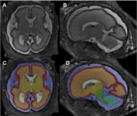
在排除了一些因脑结构异常和/或图像质量差等原因进行的胎儿MRI后,最后的研究组包括24名酒精阳性胎儿的26例胎儿MRI检查,以及52名性别和年龄匹配的健康胎儿作为对照组。在成像时,胎儿的年龄在20到37周之间。
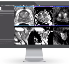
The researchers used super-resolution imaging, which allowed them to create one dataset to re-construct each fetal brain. Next, they completed an analysis of 12 different brain structures, computing total brain volume and segment volumes of specific brain compartments.
“One of the main hallmarks of our study is that we investigated so many smaller sub-compartments of the brain,” said co-author Marlene Stuempflen, M.D., scientific researcher at the Medical University of Vienna.
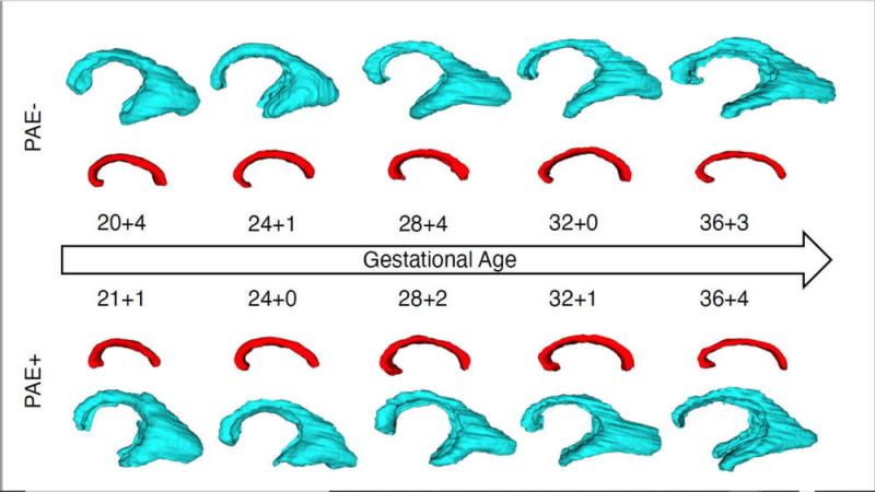
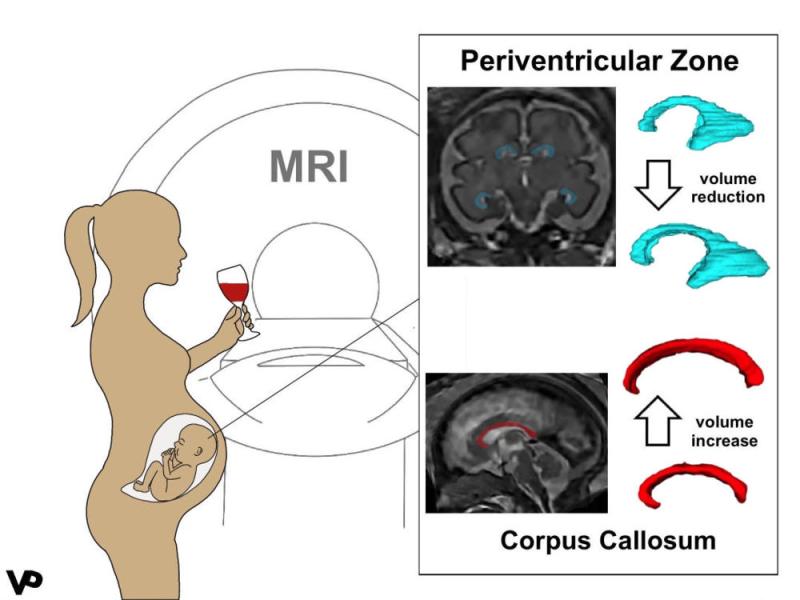
The statistical analysis revealed two major differences in the alcohol-exposed fetuses compared to healthy controls: an increased volume in the corpus collosum and a decreased volume in the periventricular zone.
“This is the first time that a prenatal imaging study has been able to quantify these early alcohol-associated changes,” Stuempflen said.
胼胝体是大脑两个半球之间的主要连接部分。Stuempflen指出,这一非常重要的结构受到影响是恰当的,因为胎儿酒精谱系障碍的临床症状是高度异质性的,或多样化的,不能准确地指出大脑的一个特定的亚结构。
“The changes found in the periventricular zone, where all neurons are born, also reflect a global effect on brain development and function,” she said.
The researchers said finding a thicker corpus collosum in the alcohol-positive fetuses was surprising because the corpus collosum is thinner in infants with fetal alcohol spectrum disorders.
卡斯普里安博士说:“看来,怀孕期间接触酒精会使大脑偏离正常的发展轨道。”“胎儿核磁共振成像是一个非常强大的工具,不仅可以表征遗传条件下的大脑发育,还可以表征因接触有毒物质而导致的后天条件。”
其他合著者有Ernst Schwartz, m.s., Mariana Diogo, m.d., Ph.D., Sarah Glatter, m.d., M.M.Sc。, Birgit Pfeiler, Victor Schmidbauer, M.D., Lisa Bartha-Doering, Ph.D., Rainer Seidl, M.D., Elisabeth Krampl-Bettelheim, M.D., and Daniela Prayer, M.D.
For more information:www.rsna.org
Additional RSNA21 conference coverage can be found here.
Related Fetal MRI Content:
COVID-19 During Pregnancy Doesn’t Harm Baby’s Brain
Researchers Generate 3-D Virtual Reality Models of Unborn Babies

 August 10, 2022