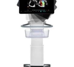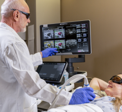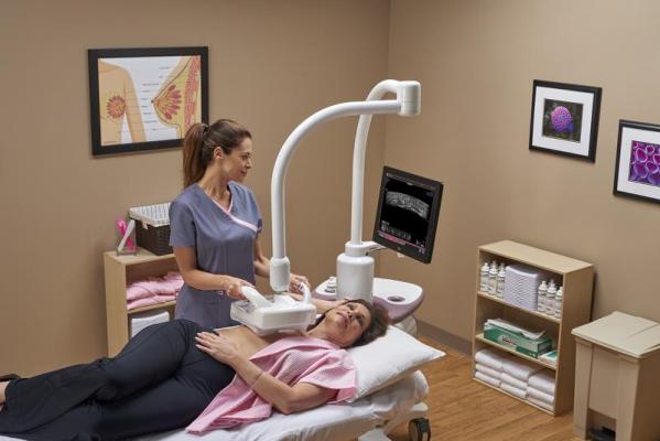
December 3, 2018 — GE Healthcare recently launched the Inveniaautomated breast ultrasound (ABUS)2.0 system in the United States. This device is the only U.S. Food and Drug Administration (FDA)-approvedultrasoundsupplemental breast screening technology, according to GE, specifically designed for detecting cancer indense breast tissue. When used in addition to mammography, Invenia ABUS can improvebreast cancerdetection by 55 percent over mammography[1]alone.[2]
“We are committed to informing patients about breast density, and offering supplemental screening options,” said Sophia Roumanis, M.D., section head of2022世界杯篮球预选赛赛程and intervention, Beaumont Breast Care Center, Dearborn, Mich. “We are thrilled to add this advanced ultrasound technology to our breast cancer screening program, which allows better visibility of dense breast tissue during breast cancer screenings.”
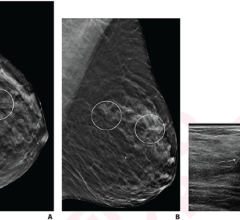
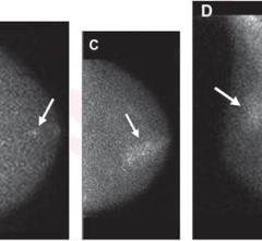
More than 40 percent of women in the U.S. have dense breast tissue[3], which is one of the strongest common risk factors for developing breast cancer. Breast density is a measurement of the amount of fatty tissue versus the amount of fibrous tissue in the breast. Both cancer and dense tissue appear white on a mammogram, so looking for tumors in women with dense breasts can be like looking for a snowball in a snowstorm. Because of this, tumors are often unseen – or “masked” – on mammography exams.
“Our goal is to find cancers as early as possible to offer the best potential outcome for the patient,” commented Vidya Pai, M.D., section head of breast imaging and intervention, Beaumont Hospital, Royal Oak, Mich. “By offering this supplemental screening to mammography for patients with dense breast tissue, we anticipate improving detection for small cancers that may not be seen on a mammogram alone in these women.”
Launched in 2014, Invenia ABUS has been installed at hundreds of facilities around the world. The new Invenia ABUS 2.0 builds on its predecessor to enhance the exam experience for both operators and patients, including new features that further customize the exam based on the patient’s body:
高效的检查:cSound Imageformer是一款基于软件的图形处理器,提供了一种可重复的、独立于操作者的采集方法,以获得一致的、高质量的结果。cSound imaging允许收集更多的数据,并用于创建每一个图像。传统的手持式超声参数如聚焦区域和增益是自动优化的。不需要对图像进行任何操作,操作人员只需按一下按钮,就能获得一致的高图像质量;and
改善患者体验:反向曲线换能器的柔和形状遵循乳房的自然轮廓,提供患者舒适,彻底接触,帮助确保全面覆盖。15厘米的大视场传感器很容易定位,在扫描时保持均匀压缩。由于没有两个女性是完全相同的,所以可以通过可编程扫描协议定制检查,可调节扫描深度和压缩水平。通过触摸按钮,一旦乳房组织采集完成,操作人员还可以缩短扫描时间。
For more information:www.gehealthcare.com
References
[1]Increase in cancer detection associated with an overall decrease in false positives.
[2]Brem RF, Tabár L, et.al. Assessing Improvement in Detection of Breast Cancer with Three-dimensional Automated Breast US in Women with Dense Breast Tissue: The SomoInsight Study. Radiology. 2015 Mar; 274(3): 663-73.
[3]https://ww5.komen.org/Breastcancer/Highbreastdensityonmammogram.html

 August 11, 2022