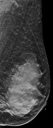
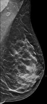
乳房密度分为四类,from lowest to highest amounts of fibroglandular tissue composition. Category A: Almost entirely fatty (least amount of fibroglandular tissue). Category B: Scattered fibroglandular tissue. Category C: Heterogeneously dense. Category D: Extremely dense (most amount of fibroglandular tissue).
Breast canceris the most common cancer in women, and, for women in their forties, the leading cause of premature death. It is the most common cause of death from cancer among Hispanic women, and the second most common cause of death from cancer among white, black, Asian/Pacific islander and American Indian/Alaska native women, according to theCenters for Disease ControlandPrevention(excluding non-melanoma skin cancers).1
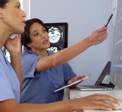
While there has been some recent controversy regarding mammography screening recommendations, theAmerican College of Radiology(ACR),Society of Breast Imaging(SBI) andAmerican Society of Breast Surgeons(ASBrS) recommend that women with an average risk of breast cancer begin getting annual screening mammograms at age 40,2,3as this is the only test that has been shown to reduce breast cancer mortality. These are read by radiologists with specialized training in breast imaging.
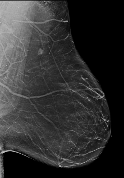
Viewing Dense Breast Tissue
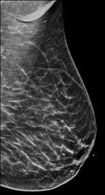
Breast tissue is composed of a mixture of milk ducts, glands and supportive tissue, which is collectively called fibroglandular tissue and fat. The radiologist who reviews the mammogram analyzes overall breast density, which is the ratio of the amount of fibroglandular tissue to fatty tissue. Women with dense breasts have more fibroglandular tissue than fatty tissue. Fibroglandular tissue appears white on a mammogram and fat appears black. It becomes harder to detect a white cancer against a white background — imagine trying to find a polar bear in a blizzard. Dense tissue is harder to see through to detect abnormal findings. As breast density increases, cancer detection becomes more difficult. Additionally, breast cancer risk rises with increasing breast density.Multiple studieshave indicated that there is a two- to six-fold increased risk of breast cancer for a woman who has an extremely dense breast versus a woman with an almost entirely fatty breast.4
乳房密度分为四类,from lowest to highest amounts of fibroglandular tissue composition:
Category A:Almost entirely fatty (least amount of fibroglandular tissue)
Category B:Scattered fibroglandular tissue
Category C:Heterogeneously dense
Category D:Extremely dense (most amount of fibroglandular tissue)
Mammography has anoverall sensitivityof 70-90 percent, which is variable, ranging from as low as 30-48 percent in patients with dense breasts to as high as 80-98 percent in patients with fatty breasts.5
Connecticut was the first state to mandate that radiologists inform all women undergoing screening mammography if they have dense breast tissue. Mammography facilities will soon be required by federal regulations to include breast density information in reports sent to patients and their physicians.
TheU.S. Food and Drug Administration(FDA) is creating standard reporting language; mammography reports and summaries must include, at a minimum, the following information:
1. How breast density may mask cancer on a mammogram.
2. A qualitative assessment of breast density as performed by the reader.
3. A reminder to individuals with dense breasts to talk with their provider if they
have questions.
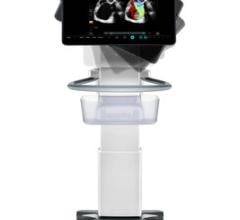
Screening Ultrasound for Women at Risk
筛查性乳房超声已被证明是一种有效的补充筛查选项,与乳房x光检查一起,对致密乳房的妇女。研究表明,超声波筛查可以提高乳腺癌的检出率。6This is due to the fact that dense breast tissue appears whiter on ultrasound, but cancers generally appear darker (unlike mammography). Often, if a woman has heterogeneously dense or extremely dense breasts, the reporting radiologist will recommend supplemental screening with breast ultrasound.
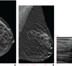
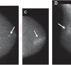
Digital breast tomosynthesis (DBT) has been shown to increase cancer detection rates compared to full field digital mammograms (FFDM) irrespective of tumor type, size or grade of cancer.7Cancers that might have remained hidden on FFDM behind overlapping dense tissue are better seen on DBT.
Depending on a patient’s lifetime risk for breast cancer, supplemental screening with breast magnetic resonance imaging (MRI) may be recommended. Recent research has indicated that screening with abbreviated breast MRI resulted in the detection of 15.2 cancers per 1,000 women versus 6.2 cancers per 1,000 women screened with digital breast tomosynthesis alone in women with dense breasts. In June 2016, New York became the first state to mandatefull insurance coverage所有筛查性乳房成像检查,包括对乳房致密的女性的补充成像(如乳房核磁共2022世界杯篮球预选赛赛程振和超声),都无需支付费用。8
Best Tools of Detection
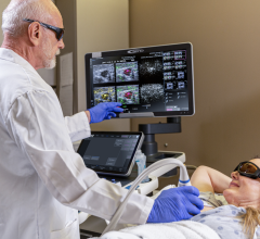
As breast imagers, our three most common tools of detection are mammography, breast ultrasound and breast MRI, which Mount Sinai Hospital uses in conjunction with the patient’s clinical history and physical exam. Early detection and treatment are the most important ways to prevent deaths from breast cancer. It is easier to successfully treat breast cancer that is detected early, when it is at a smaller size and has not spread.Regular screening tests是最可靠的方法,以早期发现和结果降低癌症分期诊断和较少需要更广泛的治疗。9Equipping women with knowledge of their breast density and personal breast cancer lifetime risk allows them to make informed decisions regarding their own most effective personalized screening regimen.
About the Authors:
Dayna Williams, M.D., is an assistant professor of radiology in breast imaging at Mount Sinai Hospital in New York, N.Y. She completed a fellowship in breast and body imaging at
Weill Cornell.
Shivani Chaudhry, M.D., is an assistant professor of radiology in breast imaging at Mount Sinai Hospital in New York, N.Y. She completedfellowships in women’s imaging at the Universityof Toronto and breast imaging
at Mount Sinai Hospital.
Laurie R Margolies, M.D., is the director of breast imaging at Mount Sinai Hospital in New York, N.Y. She is a fellow of the American College of Radiology, and was president of the New York Breast Imaging Society.
Related Dense Breast Content:
Animation to Bring Clarity to Dense Breasts
Improving Clinical Image Quality for Breast Imaging
Breast Imaging in the Age of Coronavirus
Abbreviated MRI Outperforms 3-D Mammograms at Finding Cancer in Dense Breasts
VIDEO: Use of Breast MRI Improved Cancer Detection in Dense Breasts in Dutch Study— Interview with Gillian Newstead, M.D.
Technologies to Watch in Breast Imaging
Screening MRI Detects BI-RADS 3 Breast Cancer in High-risk Patients
References:
1.https://www.cdc.gov/cancer/breast/statistics/index.htm. Accessed March 17, 2020.
2.https://www.acraccreditation.org/mammography-saves-lives/guidelines. Accessed March 17, 2020.
3.https://www.breastcancer.org/research-news/asbrs-issues-updated-screening-guidelines. Accessed March 17, 2020.
4. McCormack V, Silva I.Breast Density and Parencymal Patterns as Markers of Breast Cancer Risk: A Meta-analysis. Cancer Epidemiol Prev 2006; 15(6). June 2006.
5. Hooley RJ, Greenberg KL, Stackhouse RM, Geisel JL, Butler RS, Philpotts LE.Screening US in patients with mammographically dense breasts: initial experience with Connecticut Public Act 09-41. Radiology 2012 Oct;265(1): 59-69.
6. Berg WA, Zhang Z, Lehrer D, et al.Detection of breast cancer with addition of annual screening ultrasound or a single screening MRI to mammography in women with elevated breast cancer risk. JAMA. 2012;307(13):1394–1404. doi:10.1001/jama.2012.388.
7.https://www.sciencedaily.com/releases/2019/10/191016153705.htm
8. Comstock CE, Gatsonis C, Newstead GM, et al.Comparison of Abbreviated Breast MRI vs Digital Breast Tomosynthesis for Breast Cancer Detection Among Women With Dense Breasts Undergoing Screening. JAMA. 2020;323(8):746–756. doi:10.1001/jama.2020.0572.
9. Ahn, S., Wooster, M., Valente, C. et al.Impact of Screening Mammography on Treatment in Women Diagnosed with Breast Cancer. Ann Surg Oncol 25, 2979–2986 (2018).https://doi.org/10.1245/s10434-018-6646-8.
 August 19, 2022