
Greg Freiherr has reported on developments in radiology since 1983. He runs the consulting service, The Freiherr Group.
BLOG: Why Power Injectors Are Needed for High-Quality Imaging
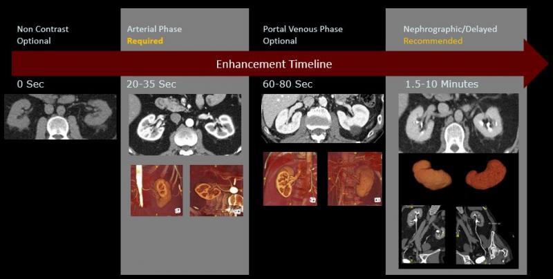
Image courtesy of Lior Molvin
造影剂改善了各种医学图像,从计算机断层扫描(CT)到磁共振成像(MR),射线摄影和透视到超声。这些检查是否具有诊断性直接取决于这些介质的适当注射。
Lior Molvin, a CT technologist and protocol manager for the diagnostic CT group atStanford Health Care, noted that nondiagnostic results waste the time of the patient, hospital staff and radiologist; and can throw a monkey wrench into scheduling, if a repeat exam is needed. They waste money and may needlessly expose the patient to radiation. Worst of all, they can critically delay treatment.
Power injectors可以直接影响图像质量和患者的安全,这位2006年开始在斯坦福大学医学中心担任技术专家的CT规程经理说。在此期间,莫尔文在医院和门诊诊断设备方面积累了丰富的经验。他的专长包括CT协议优化、技术教育和辐射安全。
The Power of the Injector
Only through the use of power injectors can contrast agents be administered at exactly the times and the flow rates needed to consistently achieve high-quality images, he said. When combined with pressure monitoring systems and hardware to prevent extravasation —definedas the accidental leakage of IV material into the surrounding tissue — these injectors can simultaneously mitigate risk.
But to be effective, technologists must consider the limitations of these machines. The jet of contrast coming from the injector can dislodge the needle from inside the vein, causing extravasation. While extravasationoccurs very infrequentlyandcomplicationsusually are minimal, complications can be severe in extreme cases.
The information to help prevent this from happening can be obtained from a manual test conducted before the CT procedure to determine whether the IV injection and the blood vessel are compatible, Molvin said. Some subtle findings might come from this test. And technologists have to be ready to act on them.
“Let’s say the protocol requires an injection of 6 mLs per second but the manual test reveals the patient would likely experience discomfort at that rate,” he said. “You could reduce the injection rate to 4mLs a second and adjust technical factors on your CT scanner to maintain image quality,” for example, the scan time and kVP.
Technologists must also consider such other issues as the concentration of the medium. High concentrations tend to produce high-quality images. “Isovue 370 always produces images that look better than 350, which look better than 320, which look better than 300,” Molvin said. “But there are times when a lower concentration is merited.”
Viscosity rises with the concentration, he explained. And high viscosities can cause extravasation, which can harm local tissue, if not properly treated.
Molvin说,一个很好的方法是调整方案,使用较低浓度的造影剂:“通过这样做,技术人员可以使用完全相同的注射参数——流速、注射体积和注射时间——然后根据患者的肾功能优化浓度。”
For example, at Stanford, a contrast medium with 300 concentration may be used instead of one with a 370 concentration, if eGFR (estimated glomerular filtration rate) indicates stage 3B chronic kidney disease, Molvin noted.
Working with Machines
People — working effectively with power injectors — are indispensible to insuring the safety of patients undergoing a contrast-enhanced CT. “There are no benefits to the patient from a nondiagnostic exam — only harm,” he said.
This harm may be physical, financial or both — physical from the unnecessary exposure of the patient to radiation; financial from the cost of the valueless exam. “A good CT scan with optimal enhancement can save the patient from having additional tests,” Molvin said.
In some cases lengthy delays could lead to death. This might happen, for example, if the CT prescribed for a patient suspected of pulmonary embolism were nondiagnostic – and the final diagnosis was delayed for too long.
“In CT you might have one shot to get it right and the contrast could make or break your study,” he said. “There (can be) incredible cost to the patient if you cut corners and don’t take good pictures.”
Greg Freiherr is a contributing editor to ITN. Over the past three decades, he has served as business and technology editor for publications in medical imaging, as well as consulted for vendors, professional organizations, academia, and financial institutions.
Editor’s Note: This is the first blog in a series titledUsing Contrast Media. The next blog will focus on how innovation can help protect patients.
Related content:
Computed Tomography Angiography: A Review and Technical Update
TH-C-18A-08: A Management Tool for CT Dose Monitoring, Analysis, and Protocol Review
Related Content

August 11, 2022 — Bracco Imaging S.p.A. has announced the renewal of the “Bracco Fellowships” initiative, in partnership ...
 August 11, 2022
2022年8月10日-迈瑞北美公司是医疗保健技术和解决方案的全球领导者和开发商……
August 10, 2022
August 10, 2022 — The imaging community has lost a legend, recognized for having revolutionized the field of diagnostic ...
August 10, 2022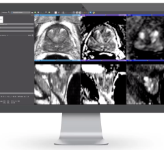
August 9, 2022 — Bot Image, an Omaha-based MRI medical device company has developed an AI-driven medical device CAD ...
August 09, 2022
August 8, 2022 — Representatives from the Diagnostics Working Group of the IASLC Early Detection and Screening Committee ...
August 08, 2022
August 5, 2022 — GE Healthcare released its most advanced fixed X-ray system yet, the next-generation Definium 656 HD ...
August 05, 2022
August 4, 2022 — Xoran Technologies announces that last month it received FDA 510(k) clearance for TRON — a truly mobile ...
August 04, 2022
August 3, 2022 — According to an open-access article in ARRS’ American Journal of Roentgenology (AJR), electronic health ...
August 03, 2022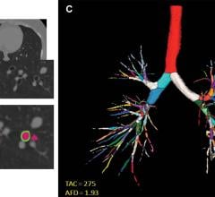
August 3, 2022 — Structural differences in lung airways between men and women may be the cause of differences in chronic ...
August 03, 2022